Short answer
- Caries is the dental term for tooth decay. A cavity becomes a root canal problem when decay stops being only a problem of the tooth's outer structure and becomes a problem of its inner tissue as well. Decay that stays in the enamel or the dentin underneath it can usually be managed by protecting or restoring the outer tooth. Decay that reaches the pulp, the soft tissue inside the tooth that contains nerves, blood vessels, and connective tissue, changes the job entirely, because the pulp itself is now inflamed or infected. At that point, saving the tooth usually means root canal treatment: the inflamed or infected pulp is removed, the inside of the tooth is cleaned and disinfected, and the space is filled and sealed. The other main option is extraction, which is removal of the tooth. The American Association of Endodontists describes those as the two main options when the nerve of a tooth is irreversibly damaged or infected. A dentist decides between them based on the condition of the tooth, the pulp, and the surrounding bone and gum, not on appearance alone. Not every cavity reaches the pulp. Many are found and treated while they are still in enamel or dentin, and the earliest stage of decay, before a hole forms, can even be stopped or reversed. The stages below show where those simpler options exist, and where they run out.
By Emma Hudson
A cavity and a root canal can sound like two unrelated problems, one small and one serious. In fact they are often two points on the same path. Decay that begins as mineral loss in the outer enamel can, if it keeps going, work inward through the dentin until it reaches the pulp, the living tissue at the center of the tooth. Once the pulp itself is inflamed or infected, the treatment has to change, because a filling repairs missing structure but does not treat tissue inside the tooth. This article explains that progression stage by stage: what is happening inside the tooth at each step, what people generally notice, what a dentist can usually do at that stage, and which warning signs mean care should not wait. It explains the process in general terms only. It does not diagnose any reader’s tooth, and no article can say how fast an individual cavity will move.
The Layers of a Tooth, in Order
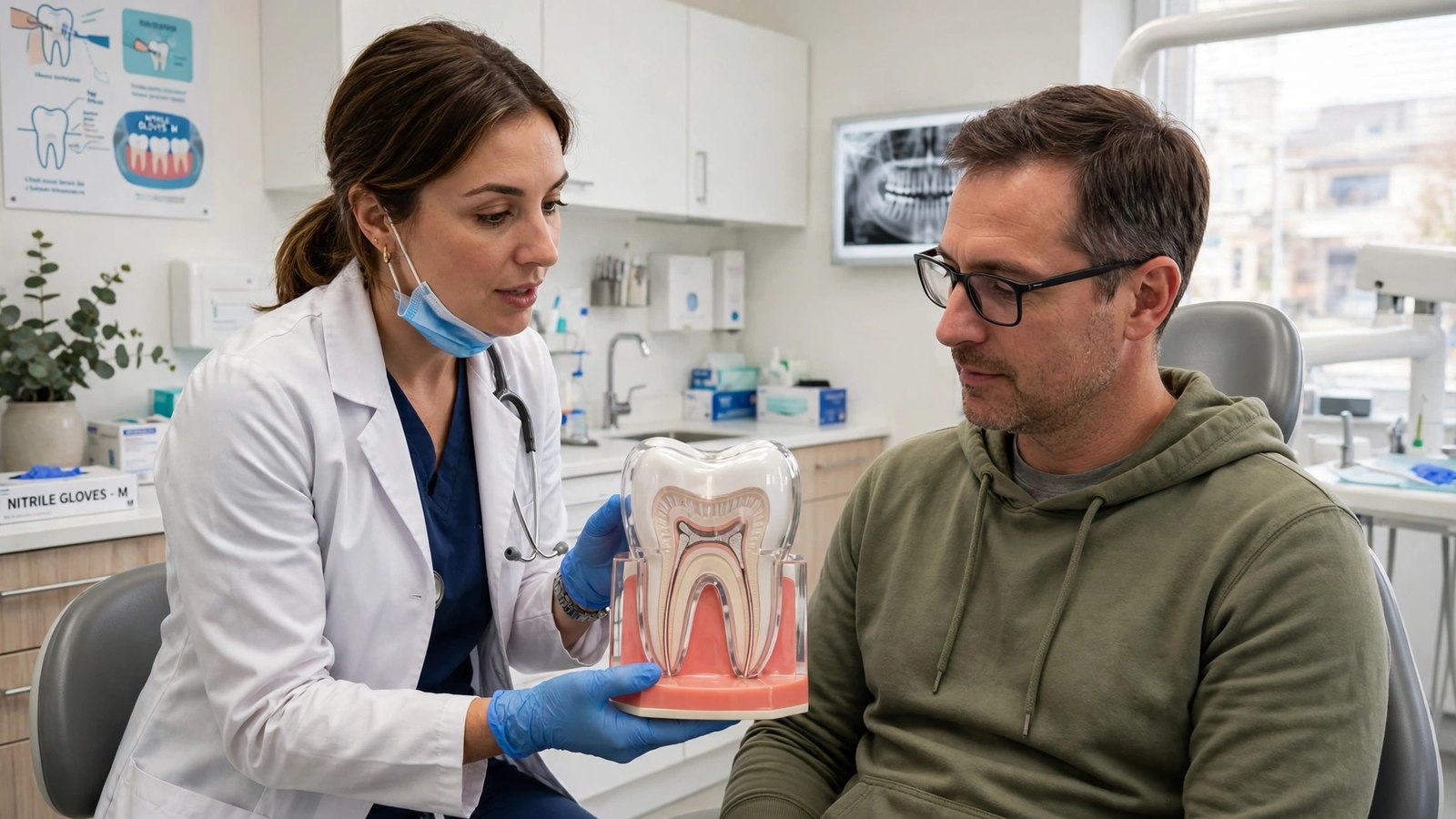
Understanding the progression means understanding the layers, because decay moves through them in order. The American Dental Association describes four dental tissues: enamel, dentin, and cementum are hard tissues, and pulp is the soft tissue at the center.
- Enamel is the hard outer layer of the crown, the part of the tooth visible above the gum. It contains no living cells and no nerves, which is why early decay in enamel alone usually causes no pain.
- Dentin is the layer beneath the enamel. It is softer and less mineralized than enamel, and it contains microscopic tubules, tiny hollow channels that lead toward the pulp. When dentin loses its enamel covering, those tubules can carry heat, cold, and sweet or acidic foods toward the nerves inside the tooth, which is one reason dentin decay often produces sensitivity.
- Pulp is the innermost living tissue. It sits in the pulp chamber in the crown and continues down the root canals to the tip of each root. It contains nerves, blood vessels, and connective tissue, and it helped build the tooth as it developed. A fully developed tooth can survive without its pulp, because surrounding tissues keep nourishing it, which is what makes root canal treatment possible.
- Cementum covers the root. It is softer than enamel, so root surfaces exposed when gums recede can decay more readily, and decay can also start at the edges of existing fillings. The starting point differs, but the inward direction of travel is the same.
Decay moves inward layer by layer, but not at a fixed speed. Diet, plaque control, fluoride exposure, saliva flow, tooth shape and position, and previous dental work all influence how quickly it advances. Any claim that a cavity will reach the pulp in a set number of months would be invented. The reliable general statement is simpler: earlier detection leaves simpler options, and deeper decay leaves fewer and more involved ones.
How Decay Starts: Acid, Mineral Loss, and Mineral Gain
Tooth decay is a process, not a single event. The National Institute of Dental and Craniofacial Research (NIDCR) explains that bacteria in the mouth make acids when they come into contact with sugars and starches from food and drink. Those acids attack the enamel and cause it to lose minerals, a process called demineralization.
At the same time, minerals in saliva, together with fluoride from toothpaste and other sources, can replace lost minerals and repair the enamel. Teeth go through this cycle of losing and regaining minerals throughout the day. Frequent eating and drinking, especially of sugary or starchy foods and drinks, tips the balance toward loss, because each exposure restarts the acid attack. Reduced saliva has a similar effect, since saliva dilutes acid, rinses the mouth, and supplies the minerals used in repair.
Which side wins decides the stage. If mineral loss is repaid, early damage heals. If loss outruns repair for long enough, the enamel weakens and eventually breaks down, and a cavity, an actual hole in the tooth, forms. That breakdown is the dividing line between damage the tooth can repair and damage it cannot. For a fuller account of what treatment can and cannot do at each point, see how to get rid of cavities: treatment options.
Stage One: Enamel Decay
The first visible sign of decay is often a white spot where minerals have been lost from the enamel. The NIDCR states plainly that tooth decay can be stopped or reversed at this point, because enamel can repair itself using minerals from saliva and fluoride from toothpaste or fluoride applied by a dentist or dental hygienist.
Treatment at this stage, in general terms, is about shifting the mineral balance back: fluoride toothpaste or professionally applied fluoride, improved cleaning, and reducing how often sugary and starchy foods and drinks reach the teeth. Some early lesions are monitored at regular exams rather than restored, because no hole has formed yet. Research into approaches that arrest or reverse early decay without restoring the tooth is covered separately in stop cavities without drilling: what new research actually shows.
Two cautions belong with the good news. First, this stage usually feels completely normal, because enamel has no nerves, so it is most often spotted at a routine exam or on an X-ray rather than noticed at home. Second, reversal has a limit. If more minerals are lost than can be restored, the enamel surface collapses into a cavity. Once a hole has formed, the missing structure does not grow back, and a restoration is usually needed to repair it. What that early damage can look like is described in what does a cavity look like.
Stage Two: Dentin Decay
Once decay breaks through the enamel, it reaches the dentin, and the character of the problem changes. Dentin is softer and more porous than enamel, so decay can spread more quickly there. It can also spread sideways under the enamel, so a cavity in dentin is often larger beneath the surface than it looks from outside. That hidden spread is one reason a spot that looks small in a mirror can need a larger restoration than expected once a dentist examines it.
Symptoms often begin at this stage, although they do not always do so. Sensitivity to cold, sweets, or pressure, or discomfort when biting, may appear as stimuli travel through the dentin tubules toward the pulp. Short sensitivity that fades as soon as the cold or sweet stimulus is gone is a different pattern from pain that lingers, and that distinction matters at the next stage. Either way, symptoms are information to report at an exam, not a diagnosis a reader can make at home.
Restoration at this stage follows how much sound tooth is left to support chewing, not how large the surface opening looks. Decay that has spread sideways can leave enamel walls too undermined to hold a filling safely, and in that situation a dentist may plan a crown, which covers and protects what remains, rather than relying on a filling alone. Where enough sound structure remains, a filling restores the tooth after the decayed tissue is removed. In selected deep cases close to the pulp, a dentist may also use a protective liner or an indirect pulp covering approach to try to keep the pulp healthy. Those are judgment calls made at the exam, based on the remaining tooth, the depth of the decay, and the condition of the pulp, and they are not available for every deep cavity.
Stage Three: Pulp Inflammation
When decay reaches the pulp, or comes close enough to irritate it severely, the pulp can become inflamed. That inflammation is called pulpitis, and dentists describe it in two forms that lead to very different treatments.
Reversible pulpitis
In reversible pulpitis, the inflammation is limited and the pulp can settle once the cause is removed. The typical pattern described in clinical references is pain or sensitivity triggered by cold or sweet stimuli that stops quickly, within seconds, once the stimulus is removed, with no spontaneous pain. Treatment aims at the cause: removing the decay and sealing the tooth with a filling, after which the pulp is expected to recover. This is the last stage at which a restoration alone can still solve the whole problem, which is why deep dentin decay is worth treating promptly rather than watching at home.
Irreversible pulpitis
In irreversible pulpitis, the inflammation will not resolve even when the cause is removed. The pattern is different: pain that lingers after hot or cold is taken away, pain that starts on its own without any stimulus, throbbing pain, or pain that wakes a person at night. Inflammation inside the rigid walls of a tooth compromises the pulp’s circulation, because there is no room for the tissue to swell. The pulp then begins to die, a process called pulp necrosis. If bacteria have entered the pulp, infection follows.
Once pulpitis is irreversible, a filling alone is no longer enough to save the tooth, because the problem is no longer only a hole in the enamel or dentin. The inflamed or infected pulp itself must be addressed. That is the turning point where a cavity problem becomes a root canal problem. It is also worth knowing that the absence of pain is not proof of safety at this stage: when the nerve dies, temperature sensitivity and spontaneous pain may fade for a time, even though infection can still be present and spreading.
Stage Four: Pulp Necrosis and Abscess
A dead pulp does not end the story. Bacteria can continue through the root canal system and out of the tip of the root into the surrounding bone. The result can be a dental abscess, a collection of pus caused by bacterial infection. The NHS describes abscess symptoms as including intense, throbbing pain that may come on suddenly and get gradually worse, pain spreading to the ear, jaw, or neck on the same side, pain that is worse when lying down, redness and swelling in the face, a tender, discolored, or loose tooth, swollen and shiny gums, sensitivity to hot or cold, and a bad taste in the mouth. If the infection spreads, a high temperature and feeling generally unwell can follow.
An abscess will not go away on its own. Dental treatment removes the source of the infection and drains the pus, which for a tooth that can be saved generally means root canal treatment, and for a tooth that cannot be saved means extraction. Antibiotics are not a substitute for that dental treatment, because they do not remove infected pulp from inside a closed tooth.
The urgent end of this stage needs to be stated clearly. In severe cases an abscess can make it hard to open the mouth fully, and can cause difficulty swallowing or breathing. The NHS treats difficulty breathing or swallowing, a lot of swelling in the mouth, and swelling affecting the eye or vision as emergencies requiring immediate emergency care. Facial swelling that is spreading, fever with tooth pain, or swelling with trouble swallowing should never be managed by waiting to see if it settles.
Why a Filling Cannot Fix Pulp Infection
It helps to think of a tooth with pulp involvement as having two separate jobs that need doing. One job is on the outside: restoring shape and strength where structure is missing. The other job is on the inside: dealing with living tissue that is inflamed or infected inside a closed chamber. A filling does the first job well. It does not reach the second job at all, because it sits in the outer tooth while the pulp chamber and root canals remain untouched.
Leaving the inner job undone matters because infection inside a tooth can continue to cause pain, swelling, bone damage around the root tip, or an abscess, even while the outside of the tooth looks repaired. That is why the treatment plan changes once the pulp is involved, even though the tooth may look similar from outside to a tooth with a deep filling.
The same logic explains why waiting changes the menu of options. A cavity that could have been filled at the dentin stage may need a crown once more structure is lost, and a tooth that could have been crowned may need root canal treatment once the pulp is involved. A tooth whose pulp infection has destroyed too much structure, or cracked it beyond repair, may leave extraction as the only option. Each step involves deeper tissue or removes more tooth, and each step is more involved than the last. Early exams break that chain by finding decay while the simpler options remain.
What Happens During Root Canal Treatment
Root canal treatment, also called endodontic treatment, is planned when a tooth is worth keeping but its pulp cannot stay. The American Association of Endodontists summarizes the procedure as removing the inflamed or infected pulp, carefully cleaning and disinfecting the inside of the tooth, then filling and sealing it, which eliminates bacteria from the canal, helps prevent reinfection, and saves the natural tooth. In general terms, the steps are:
- Examination and imaging. The dentist examines the tooth and takes X-rays to see the shape of the root canals and check for signs of infection in the surrounding bone.
- Numbing and isolation. Local anesthetic is given to numb the area. A protective sheet, often called a rubber dam, is placed around the tooth to keep it dry and isolated from saliva while the work is done.
- Access. A small opening is made through the crown of the tooth to reach the pulp chamber, and any decay in the way is removed.
- Pulp removal and cleaning. The inflamed or infected pulp is removed from the chamber and the root canals. The canals are cleaned, shaped, and disinfected with irrigating solutions so the sealing material can fill them properly.
- Filling and sealing. The cleaned canals are filled and sealed, commonly with a rubber-like material called gutta-percha, together with a sealing cement. If treatment takes more than one visit, a temporary filling may protect the tooth in between, and medication may be placed inside to help eliminate bacteria.
- Final restoration. The tooth is rebuilt with a filling or, commonly for a back tooth, a crown. This step is part of the treatment, not an optional extra, because the tooth has usually already lost a lot of structure and a treated tooth can be more prone to fracture under chewing forces.
Treatment is carried out by a general dentist or by an endodontist, a specialist with additional training in treating the inside of teeth, to whom complex cases are often referred. The number of visits varies with the tooth and the infection. No outcome can be guaranteed for any individual tooth, and a treated tooth still needs the same daily care as any other, because its outer structure can still decay. Practical care after fillings, crowns, and other restorative work is covered in crown, bridge, and filling aftercare.
The Alternatives When Pulp Is Involved
Root canal treatment is not the only path, and it is not right for every tooth. The realistic alternatives are:
- Extraction. Removing the tooth removes the infection source with it. It is the option when the tooth is too broken down, cracked, or unsupported by bone and gum to restore reliably. Extraction leaves a gap, and replacing the tooth with a bridge, an implant, or a partial denture is a separate decision with its own costs and treatment steps, to discuss with a dentist. Leaving the gap is also a decision, and it can affect chewing and the position of neighboring teeth over time.
- Saving the tooth with root canal treatment. Keeping the natural tooth preserves normal biting and chewing, keeps neighboring teeth in their positions, and avoids replacement treatment. The American Association of Endodontists notes that this preservation is the major advantage of root canal treatment over extraction and replacement.
- Doing nothing. This is sometimes chosen by default rather than by decision, and it is the option with the clearest downside: untreated pulp infection can continue to cause pain or lead to an abscess, and the tooth may be lost anyway, under worse conditions. Pain that fades because the nerve has died is not the infection resolving.
One further distinction matters. Antibiotics and pain relief can have a supporting role when infection has spread beyond the tooth or when there are general symptoms such as fever, as a dentist or doctor judges. Neither removes the infected pulp, so neither is a standalone treatment for pulp infection.
Prevention at Each Stage
Prevention works best when it is matched to the stage it is trying to stop.
Before decay starts, and at the white spot stage. Brush twice a day with fluoride toothpaste and clean between the teeth daily, which reduces the plaque that produces acid. Fluoride prevents mineral loss and replaces lost minerals, which is why it can stop or reverse early decay before a hole forms. Limiting how often sugary and starchy foods and drinks are consumed reduces the number of acid attacks each day, and keeping regular dental appointments gives a dentist the chance to spot white spots while reversal is still possible. Anyone whose mouth is persistently dry, whether from medication or another cause, should mention it at an exam, because reduced saliva removes one of the tooth’s main defenses.
On chewing surfaces. Dental sealants are thin protective coatings applied to the chewing surfaces of back teeth, where deep grooves trap plaque and decay often starts. The Centers for Disease Control and Prevention (CDC) reports that two years after placement, sealants prevent more than 80 percent of cavities in permanent molars, and that school-aged children without sealants had almost three times more cavities in permanent first molars than children with sealants. Sealants are mostly discussed for children and teens, whose molars are newly erupted, and a dentist can advise whether they suit a particular patient.
At the dentin stage. Prevention here means not postponing the restoration. A cavity that needs a filling will not heal under better brushing, because the hole itself is permanent damage. Keeping the appointment, and reporting new sensitivity in a filled tooth rather than waiting for it to worsen, protects the pulp while reversible treatment is still available.
After root canal treatment. A treated tooth can still develop new decay around the edges of a filling or crown, and gum disease can still affect it. Completing the final crown or restoration on schedule, then continuing brushing, cleaning between teeth, and regular exams, is what protects the investment the treatment represents.
When to Seek Care Promptly
See a dentist promptly if you have severe tooth pain, pain that lingers after hot or cold is removed, pain that starts on its own or wakes you at night, swelling in the gums, cheek, or jaw, fever together with tooth pain, pus or drainage near a tooth, a small bump on the gum near a painful tooth, a bad taste together with pain or swelling, or pain when biting that is getting worse. Do not try to treat pulp infection at home, and do not use pain relief alone to decide whether care is needed, because pain relief can mask symptoms while infection continues, and pain can also fade because the pulp has died while infection remains.
Seek emergency medical care the same day if you have trouble breathing, trouble swallowing, swelling that is spreading across the face or toward the eye, difficulty opening your mouth, or a high temperature with facial swelling and feeling very unwell. Those are signs that infection may be spreading beyond the tooth, and they outrank a routine dental appointment.
Two history points are worth mentioning at any exam. A previously filled tooth that now hurts, and a tooth with a deep cavity that now has lingering sensitivity to hot or cold, both deserve assessment. Previous treatment, cracks, and repeated dental work on the same tooth can also affect the pulp, even without new decay, so a dentist weighs decay together with the tooth’s whole history.
Frequently Asked Questions
When do caries lead to root canals?
Caries lead to root canals when decay reaches the pulp and the pulp becomes irreversibly inflamed or infected, so the problem is no longer only a hole in the outer tooth. At that point the pulp cannot recover even if the decay is removed, and saving the tooth means removing the pulp and sealing the inside, which is root canal treatment. Not every cavity reaches that point. Early detection and treatment can keep decay at the enamel or dentin stage, where protection or restoration alone can still work.
Does every deep cavity need a root canal?
No. A deep cavity needs an exam to judge how close the decay is to the pulp and whether the pulp is reversibly or irreversibly inflamed. Some deep cavities can be treated with a larger restoration or a crown, sometimes with a protective covering over the pulp. Others need root canal treatment. Appearance alone cannot decide, and neither can pain alone, because some infected teeth hurt very little. The dentist’s examination, pulp tests, and X-rays decide together.
How long does it take for a cavity to reach the pulp?
No safe timeline can be given for an individual tooth. Progression speed varies with diet and how often teeth meet sugar and starch, plaque control, fluoride exposure, saliva flow, tooth location and shape, and previous dental work. That variability is exactly why dentists advise against waiting for pain to make the decision: enamel decay is painless, dentin decay is inconsistent, and by the time pain is reliable the pulp is usually already involved.
Is root canal treatment the only option for pulp infection?
No. The main options are root canal treatment to save the tooth, or extraction to remove it. A dentist decides based on how much sound tooth remains, the condition of the pulp and root, the surrounding bone and gum, and the overall treatment plan. Antibiotics and pain relief may support treatment when infection has spread, but neither one removes infected pulp, so neither replaces one of those two options.
Can a tooth that has had a root canal still get a cavity?
Yes. Root canal treatment removes the pulp, not the outer tooth. The enamel, dentin, and the edges of any filling or crown can still decay, and gum disease can still affect the tooth and its support. That is why the final restoration matters, why aftercare matters, and why brushing, cleaning between teeth, and regular exams remain just as important for a treated tooth as for any other. See crown, bridge, and filling aftercare for the practical detail.
Does root canal treatment hurt?
The procedure is done under local anesthetic, and guidance from dental organizations describes it as a routine treatment intended to relieve the pain of pulp infection rather than cause pain. The pain most people associate with root canals is usually the pain of the infected or irreversibly inflamed pulp beforehand. Some tenderness for a few days afterward is common as the tissues around the root settle. A specific experience cannot be promised for any individual, and worsening pain or new swelling after treatment should be reported to the treating dentist promptly.
Sources & references
- National Institute of Dental and Craniofacial Research (NIDCR), "Tooth Decay": https://www.nidcr.nih.gov/health-info/tooth-decay
- National Institute of Dental and Craniofacial Research (NIDCR), "The Tooth Decay Process: How to Reverse It and Avoid a Cavity": https://www.nidcr.nih.gov/health-info/tooth-decay/more-info/tooth-decay-process
- American Dental Association, MouthHealthy, "Tooth": https://www.mouthhealthy.org/all-topics-a-z/tooth.aspx/
- Journal of the American Dental Association, "Getting to the root of endodontic (root canal) treatments": https://jada.ada.org/article/S0002-8177(14)61651-2/fulltext
- American Association of Endodontists, "What is a Root Canal?": https://www.aae.org/patients/root-canal-treatment/what-is-a-root-canal/
- American Association of Endodontists, "Root Canal vs. Extraction": https://www.aae.org/patients/root-canal-treatment/what-is-a-root-canal/root-canal-vs-extraction/
- NHS 111 Wales, "Root Canal Treatment": https://111.wales.nhs.uk/rootcanaltreatment/
- NHS Inform, "Dental Abscess": https://www.nhsinform.scot/illnesses-and-conditions/mouth/dental-abscess/
- Centers for Disease Control and Prevention, "Vital Signs: Dental Sealant Use and Untreated Tooth Decay Among U.S. School-Aged Children", MMWR: https://www.cdc.gov/mmwr/volumes/65/wr/mm6541e1.htm
- Journal of the Canadian Dental Association, "Pulpitis (Reversible/Irreversible)": https://jcda.ca/node/13726


