Short answer
- Your heart sits in the center of your chest, in a compartment called the mediastinum, tucked between your two lungs and behind the breastbone.
- It is tilted, not left-sided. Roughly two-thirds of its mass lies to the left of the midline and one-third to the right, the standard description in anatomy textbooks.
- The heart's broad top, the base, sits high in the chest. Its pointed lower tip, the apex, points down and to the left, ending just left of the breastbone between the fourth and fifth ribs.
- You feel your heartbeat on the left because the apex is the part that presses closest to the chest wall with each beat, and the thick-walled left ventricle does the strongest pumping.
- The position is set before birth, when the primitive heart tube folds into an S shape during the fourth week of development.
- In about 1 in 12,000 pregnancies the heart develops on the right instead, a variation called dextrocardia. In about 1 in 10,000 people, all the internal organs are mirrored, a pattern called situs inversus totalis.
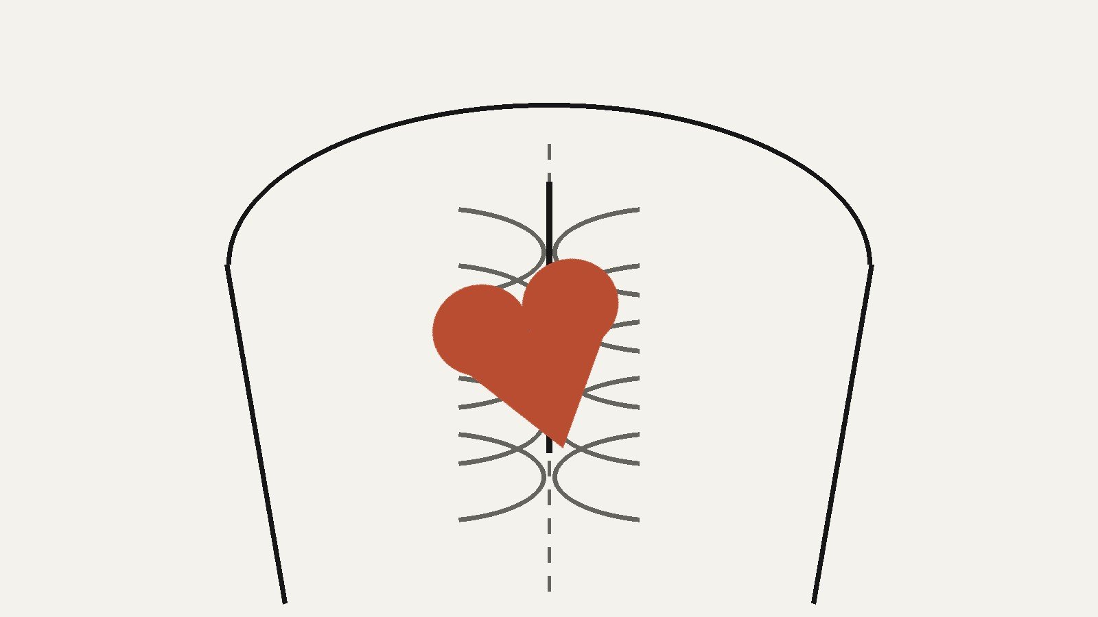
Place a hand flat on your chest where you expect your heart to be, and you will almost certainly put it high on the left. Schoolchildren do it for pledges, actors do it in hospital scenes, and most adults asked to point at their heart aim at the same spot. The gesture is close, but it is not accurate. Your heart is not a left-side organ. It sits in the center of your chest, behind your breastbone, and leans. About two-thirds of it lies to the left of your body’s midline and about one-third to the right, and its lower tip angles down and to the left, which is why the beat you feel is a left-side event. This article explains the position properly: where the heart sits, why the leftward lean exists, how it is established before birth, and the rare people in whom the whole arrangement is mirrored.
This article explains anatomy only, and it is not medical guidance: chest pain or pressure is an emergency that needs immediate medical care.
The Precise Answer: Center, Tilted Left
OpenStax’s Anatomy and Physiology, the textbook used across introductory anatomy courses, puts it this way: “The human heart is located within the thoracic cavity, medially between the lungs in the space known as the mediastinum.” Medially means toward the midline. The heart is a central organ, resting directly behind the sternum, the flat bone in the middle of your chest, with the lungs on either side of it and the diaphragm, the sheet of muscle under the lungs, beneath it.
Central does not mean symmetrical. The heart is a roughly cone-shaped organ about the size of your fist, and it sits rotated. Its wide end, the base, is at the top, positioned at about the level of the third costal cartilage, the gristly joint where the third rib meets the breastbone. From there the organ runs downward and to the left, ending in the apex, the blunt tip that OpenStax locates “just to the left of the sternum between the junction of the fourth and fifth ribs.” Because of that diagonal run, the bulk of the organ ends up left of center even though it starts in the middle: about two-thirds of its mass to the left of the midsternal line, one-third to the right.
The rotation goes one step further. The right side of the heart is turned toward the front of the chest and the left side toward the back, so the chambers are not arranged in a tidy left-right row. The lung on the left even makes room: the medial surface of the left lung carries a depression called the cardiac notch, a literal dent where the heart’s leftward lean presses in. The left lung, partly for this reason, has two lobes to the right lung’s three.
So the one-line answer to the title question is: the left, mostly, but only because a central organ leans that way. “Center, tilted left” is the version an anatomist would sign.
Why Everyone Says “Left Side”
If the heart is central, why is the left-side belief so universal? Because the evidence of your own senses points there.
The beat is a left-side event. Each contraction drives the apex forward against the chest wall. The spot where that impact is strongest, clinicians call it the point of maximal impulse, sits on the left, around where the apex ends. Put a palm on your chest and the thump you feel is the apex arriving, not the whole heart.
The strong chamber is on the left. The left ventricle, which pumps blood to the entire body rather than just to the nearby lungs, has by far the thickest wall of the four chambers and sits on the heart’s left and rear. The hardest-working, hardest-thumping part of the organ is leftward, so its sounds and sensations are too. A stethoscope placed to hear the heart at its clearest goes over the apex, on the left.
Culture rehearsed the gesture. Portraits, films, pledges and Valentine iconography all put the heart on the left, so the idea is practiced physically thousands of times before anyone meets an anatomy diagram. The icon is not even wrong about where the beat is. It just mistakes the loudest part of the organ for the whole of it.
Base and Apex: The Two Ends, and a Naming Trap
Anatomy describes the heart’s position using its two ends, and the names confuse everyone the first time, because the base is at the top.
The base is the heart’s broad upper surface. This is where the great vessels attach: the superior and inferior venae cavae bringing blood back from the body, and the aorta and pulmonary trunk carrying blood away. OpenStax places the base at the level of the third costal cartilage, high behind the breastbone. Its position near the top, close to the back of the chest, is why anatomists call it the base: it is the anchored end, fixed by the vessels entering and leaving it.
The apex is the opposite end, the rounded tip formed by the left ventricle, pointing down, forward and to the left. It is the mobile end, the one that swings against the chest wall with each beat. When this article and every textbook say the heart “points left,” it is the apex doing the pointing.
Holding both facts at once resolves most confusion. The heart’s top is central and high. Its bottom tip is left and low. Describing the whole organ by the tip’s address, “on the left,” throws away the top two-thirds of the story.
The Pericardium: The Sac That Holds It in Place
The heart does not float loose in the chest. Within the mediastinum it sits inside the pericardium, a tough double-layered sac that OpenStax describes as separating the heart from the other structures of the mediastinum and giving it its own space, the pericardial cavity.
The outer layer, the fibrous pericardium, is dense connective tissue anchored to the diaphragm below and to the great vessels above, which is part of why the heart’s position is so consistent from person to person. Inside it, the serous pericardium has two layers of its own: a parietal layer lining the fibrous sac, and a visceral layer, also called the epicardium, that is the heart’s own outer surface. Between them is a thin film of lubricating fluid, letting the heart beat roughly 100,000 times a day against its neighbors without friction wear.
The pericardium also limits how far the heart can shift or overfill, which is the mechanical reason the heart stays where the developmental process puts it, tilted in the center, for life.
Set Before Birth: How the Heart Tube Folds Left
The heart is the first organ to become functional in a developing embryo, and its leftward lean is one of the earliest left-right decisions the body makes.
In the third week of development, two small vessels called endocardial tubes fuse in the midline to form a single primitive heart tube. At that stage the heart is a straightish tube, with blood entering at one end and leaving at the other, and no left lean at all. Then, as OpenStax’s development chapter describes, the tube elongates faster than the space containing it and “begins to fold within the pericardium, eventually forming an S shape, which places the chambers and major vessels into an alignment similar to the adult heart.” That folding happens between days 23 and 28.
The fold is not decorative. The tube’s segments become specific structures: the bulbus cordis develops into the right ventricle, the primitive ventricle forms the left ventricle, and the primitive atrium becomes the front portions of both atria. Because the loop bends the way it does, the ventricular end of the tube, the future apex, is carried down and to the left, while the atrial end settles up and behind. The adult arrangement, base high and central, apex low and left, is the folded tube’s geometry preserved.
The medical literature on the variation in the next section describes the same step from the other side: looping determines the position of the ventricles relative to the atria, and when the looping and migration go the other way, the heart can end up in the right half of the chest. Position, in other words, is not something the heart settles into after birth. It is a folding decision made in the fourth week, and it is normally made the same way in almost everyone.
The Mirror Image: Dextrocardia and Situs Inversus
In a small minority of people, the fold, and sometimes the whole body’s left-right pattern, runs in reverse.
Dextrocardia means the heart sits in the right side of the chest with its apex pointing right. StatPearls, the clinical reference published through the National Library of Medicine’s Bookshelf, calls it “a very rare condition,” with studies finding incidence rates of “around 1 in 12,000 pregnancies.” It arises during that same looping stage: if the developing heart fails to migrate into the left half of the chest, or completes its rotation on the right, the result is a right-sided heart. Dextrocardia by itself is a positional fact, not a verdict on health, and because a mirrored heart can work normally, it is frequently discovered by accident, when a chest image taken for some unrelated reason shows the heart on the unexpected side.
Situs inversus totalis is the fuller mirror: all of the internal organs are reversed left for right, so the heart is on the right, the liver on the left, the stomach and spleen on the right, each in the reflected position. Published estimates put it at roughly 1 in 10,000 people, with study figures ranging from about 1 in 8,000 to 1 in 25,000. Where dextrocardia occurs as part of situs inversus, the organs are at least consistently mirrored, a complete set facing the other way.
Between the usual pattern and the full mirror sit rarer mixed arrangements, grouped under the term heterotaxy, in which some organs are reversed and others are not. Those are considerably less common than either pattern above, and considerably more complicated; they belong to specialist literature rather than to a position explainer, and they are mentioned here only so the map of possibilities is complete.
One caution applies to everything in this section: these are population figures about anatomy, not information about any individual reader. A heart position is something established by imaging, not something to reason about from sensations at home.
Part of a Wider Left-Right Pattern
The heart’s lean looks less like an oddity once you notice that the body is asymmetric throughout, in a pattern anatomists call situs solitus, the usual arrangement.
The liver, the body’s largest internal organ, sits mostly under the right ribs. The stomach and the spleen sit on the left. The right lung is broader and has three lobes; the left lung is narrower, carries the cardiac notch, and has two. Even the two sides of the heart are unequal partners, the left ventricle built far thicker than the right. Left and right in the body are not mirror halves with identical contents. They are two different neighborhoods, and the heart leaning left is one feature of a body-wide layout that the embryonic folding establishes all at once.
This is also why the mirror conditions in the previous section are described the way they are: situs is the name for the whole pattern, not for any single organ’s address.
How Doctors Describe Position
Medical descriptions of the heart sound more left-sided than this article’s answer, and the difference is mostly about reference frames.
Descriptions assume the anatomical position: the subject standing upright, facing the observer, arms at the sides. Left and right in an anatomy text or a medical report always mean the subject’s left and right, never the viewer’s. Medical images follow the matching convention: a chest image is displayed as if you were facing the patient, so the patient’s left appears on your right as you look at the screen. Once you know that, an apparently right-sided heart on a displayed scan is usually the normal left-leaning heart, correctly presented.
Clinicians also locate the heart against surface landmarks rather than sides: the base at the level of the third costal cartilage, the apex just left of the sternum between the fourth and fifth ribs. Those coordinates, not “left,” are what a precise position statement looks like, and they describe a central organ with a leftward tip, exactly as the anatomy says.
Frequently Asked Questions
Is the heart on the left or the right side of the body?
Neither, exactly. It sits in the center of the chest, in the mediastinum behind the breastbone, and leans: about two-thirds of its mass lies left of the midline and one-third right, with its tip, the apex, pointing down and to the left. “On the left” is a serviceable shorthand and a poor description.
Why can I feel my heartbeat on the left?
Because the apex, the heart’s tip, ends on the left, just inside the chest wall between the fourth and fifth ribs, and each beat pushes it against that wall. The thick-walled left ventricle, the strongest chamber, forms the apex, which is why the impact and the clearest heart sounds are both found on the left.
Do some people really have their heart on the right?
Yes. The variation is called dextrocardia, and clinical references put it at around 1 in 12,000 pregnancies. In some of those people the heart alone is right-sided; in others it is part of situs inversus totalis, a complete mirror image of the internal organs found in roughly 1 in 10,000 people. It is often discovered incidentally on imaging done for other reasons.
Is the heart in exactly the same place in everyone?
The pattern is remarkably consistent: central chest, base high, apex low and left, held in place by the pericardium and its attachments. Small differences in body build and chest shape shift the exact coordinates a little, and the heart moves slightly with breathing and posture, but the arrangement itself is a standard feature of human anatomy, set by the folding of the heart tube before birth.
Why is the top of the heart called the base?
Because it is the anchored, vessel-bearing end. The great veins and arteries attach at the heart’s broad upper surface, which sits high in the chest near the level of the third costal cartilage, while the free lower tip is the apex. The naming follows the organ’s fixed and free ends, not the direction you might expect from the word.
Conclusion
Your heart is a central organ with a leftward lean: seated in the mediastinum between the lungs, held in its pericardial sac behind the breastbone, about two-thirds of it left of the midline, its apex tipped down and to the left where you can feel it beat. The position is not an accident of adult anatomy but a folding pattern completed in the fourth week of development, repeated in almost every human body, and reversed, heart alone or with every organ, in a small and well-documented minority. “Left side” survives as an answer because the beat is on the left. The heart itself is in the middle, leaning.
Keep reading: The Human Skeleton: Structure, Divisions and Functions
Sources & references
- OpenStax (Rice University), Anatomy and Physiology 2e, "Heart Anatomy," Location of the Heart: the heart is located within the thoracic cavity, medially between the lungs in the mediastinum; it is separated from the other mediastinal structures by the pericardium and sits in the pericardial cavity; the base is at the level of the third costal cartilage; the apex lies just to the left of the sternum between the junction of the fourth and fifth ribs; the right side of the heart is deflected anteriorly and the left side posteriorly; the cardiac notch of the left lung reflects the apex's deviation.
- OpenStax (Rice University), Anatomy and Physiology 2e, "Development of the Heart": the primitive heart tube elongates and folds within the pericardium into an S shape between days 23 and 28, aligning the chambers and great vessels as in the adult heart; the bulbus cordis develops into the right ventricle and the primitive ventricle forms the left ventricle.
- Nair R. and Muthukuru S.R., "Dextrocardia," StatPearls, National Center for Biotechnology Information (NCBI) Bookshelf, National Library of Medicine: the heart is the first organ to develop; looping of the heart tube determines the position of the ventricles relative to the atria, and failure of the loop to migrate into the left hemithorax results in dextrocardia; incidence of dextrocardia is around 1 in 12,000 pregnancies.
- MedlinePlus, U.S. National Library of Medicine: chest pain guidance (chest pain or pressure is an emergency symptom requiring immediate medical care) and encyclopedia material on situs inversus as a mirror-image arrangement of the internal organs; published estimates for situs inversus totalis center on roughly 1 in 10,000 people, with study figures ranging from about 1 in 8,000 to 1 in 25,000.
- Standard anatomy textbook description (as summarized in the Cleveland Clinic Cardiology Board Review and introductory anatomy references): the heart lies within the middle mediastinum with about two-thirds of its mass to the left of the midsternal line and one-third to the right.


