Short answer
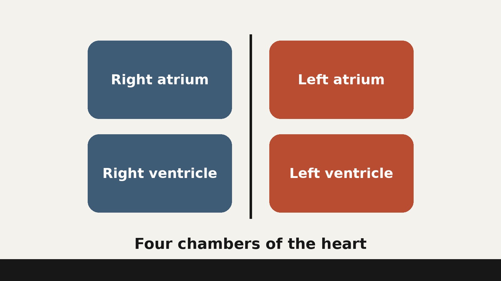
- The human heart has four chambers: two upper chambers called atria (singular: atrium) and two lower chambers called ventricles. The right atrium and right ventricle form the right side of the heart, and the left atrium and left ventricle form the left side. The atria receive blood as it arrives at the heart, and the ventricles pump blood out of the heart, either to the lungs or to the rest of the body. Valves between the chambers keep blood moving in one direction, and a muscular wall called the septum keeps the right and left sides separate. This article explains anatomy only and is not medical guidance. --- If you have ever seen a diagram of the heart, you have seen the chambers, even if you did not know their names. They are the four hollow spaces inside the organ, divided by walls and guarded by valves, through which every drop of blood passes on its round trip. The question behind this article sounds simple, "how many chambers are in the heart," but a full answer needs more than a number: the name of each chamber, what flows into it and out of it, and why the heart is built this way rather than as a single hollow bag.
What a Chamber Actually Is
A chamber of the heart is not a room with still air in it. It is a hollow space enclosed by heart muscle, lined with a smooth inner layer, that fills with blood and then empties by contracting. Think of it less like a room in a house and more like a squeezable pouch built into a pump: blood flows in, the muscular wall tightens, and blood is pushed onward to the next stage.
All four chambers share that design, but they are not interchangeable. The two upper chambers, the atria, are receiving chambers. Their main job is to collect blood that is already flowing toward the heart and pass it down into the ventricles. The two lower chambers, the ventricles, are the pumping chambers. Their walls are thicker and stronger, because they must generate enough pressure to drive blood through the lungs or through the entire body.
That division of labor is the key to the whole organ. A receiving chamber can be thin walled and flexible, because blood arrives under relatively low pressure and mostly needs somewhere to go. A pumping chamber must be thick walled and powerful, because it has to push blood onward against resistance. The heart gives each job its own chamber, on each side of the body, so four chambers in total.
One naming point causes confusion in diagrams. When you look at a picture of someone else’s heart, their right side appears on the left side of the picture, because you are facing them. Anatomists always name the chambers from the point of view of the person whose heart it is.
The Four Chambers at a Glance
| Chamber | Position | Receives blood from | Pumps blood to | Main job |
|---|---|---|---|---|
| Right atrium | Upper right | Body, through the superior and inferior venae cavae, plus the coronary sinus | Right ventricle, through the tricuspid valve | Collect blood returning from the body |
| Right ventricle | Lower right | Right atrium, through the tricuspid valve | Lungs, through the pulmonary trunk and pulmonary arteries | Pump blood to the lungs |
| Left atrium | Upper left | Lungs, through the four pulmonary veins | Left ventricle, through the mitral valve | Collect blood returning from the lungs |
| Left ventricle | Lower left | Left atrium, through the mitral valve | Body, through the aorta | Pump blood to the whole body |
Keep this table in mind for the chamber by chamber tour that follows: four chambers, two for receiving, two for pumping, split evenly between the right and left sides.
The Right Atrium: Where Blood from the Body Arrives
The right atrium is the receiving chamber for blood returning from the body. Two large veins deliver most of it. The superior vena cava drains blood from regions above the diaphragm: the head, neck, upper limbs, and the chest region. The inferior vena cava drains blood from regions below the diaphragm: the lower limbs and the abdominal and pelvic region. Both empty into the back of the right atrium, the superior vena cava above and the inferior vena cava below it. A third, smaller vessel, the coronary sinus, drains blood from the heart muscle itself into the right atrium as well.
The blood arriving here is relatively low in oxygen, because the body’s tissues have already taken much of the oxygen from it and loaded it with carbon dioxide in exchange. The right atrium simply collects it and passes it on. Blood flows into the atria nearly continuously: the atria keep accepting blood even while the ventricles below them are contracting, so the flow back toward the heart never has to stop and wait.
Inside, the right atrium is mostly smooth walled, with ridges of muscle called pectinate muscles on its front surface and a shallow oval depression, the fossa ovalis, on the wall between the two atria (see the septa section below). It passes blood down into the right ventricle through an opening guarded by the tricuspid valve. Most filling of the ventricle happens while the atrium is relaxed; just before the ventricle contracts, the atrium adds a final contractile push, topping it up.
The Right Ventricle: The Pump to the Lungs
The right ventricle receives blood from the right atrium through the tricuspid valve, and its job is to send that blood to the lungs. When it contracts, it pushes blood into the pulmonary trunk, a large vessel that soon splits into the left and right pulmonary arteries, one for each lung. In the tiny capillaries of the lungs, carbon dioxide leaves the blood and oxygen enters it. That gas exchange is the whole reason the right side of the heart exists: it is a dedicated delivery service to the lungs, so blood can be refreshed before it goes back out to the body.
The right ventricle is a pumping chamber, so its wall is thicker than an atrium’s wall, but much thinner than the left ventricle’s wall. The reason is distance and resistance: the lungs are close by, and the pulmonary circuit is short with relatively low resistance. Although both ventricles pump the same amount of blood with each contraction, the left ventricle must generate far greater pressure for the long systemic circuit, so its muscle is much thicker and better developed.
Inside the right ventricle, the wall is lined with ridges of cardiac muscle called trabeculae carneae, plus a band of muscle called the moderator band and three papillary muscles that anchor the cords of the tricuspid valve. At the exit sits the pulmonary valve, at the base of the pulmonary trunk, which lets blood leave for the lungs and stops it flowing back when the ventricle relaxes.
The Left Atrium: Where Blood from the Lungs Arrives
The left atrium mirrors the right atrium, but for the other circuit. It is the receiving chamber for blood coming back from the lungs, and that blood is now high in oxygen. It arrives through the four pulmonary veins, which carry blood from the pulmonary capillaries back to the heart. These veins are a famous exception to a general rule: veins usually carry blood low in oxygen toward the heart, but the pulmonary veins carry blood high in oxygen toward the heart, because they are returning from the place where oxygen was just picked up.
Like the right atrium, the left atrium receives blood nearly continuously. Blood flows from the pulmonary veins into the atrium and then, when the ventricle below is relaxed, onward into the left ventricle through an opening guarded by the mitral valve, also called the bicuspid valve. The left atrium does not have pectinate muscles across its main wall, though its small ear-shaped extension, the auricle, does contain such ridges. Auricles, sometimes called atrial appendages, can fill with blood and empty back into the atria.
The left atrium’s job sounds quiet, collect and pass on, but it is essential: without it, blood returning from the lungs under low pressure would have nowhere to gather while the left ventricle is contracting.
The Left Ventricle: The Body’s Main Pump
The left ventricle is the strongest chamber in the heart and the main pump for the entire body. It receives blood high in oxygen from the left atrium through the mitral valve, and when it contracts it ejects that blood into the aorta, the largest artery in the body, through the aortic valve. From the aorta, blood branches out to every tissue: the brain, the arms, the abdominal organs, the legs, and back again.
The wall of the left ventricle is the thickest of the four chambers by a wide margin, and the reason is workload. The systemic circuit, the loop through the whole body, is long and offers high resistance, so the left ventricle must generate great pressure to push blood through it. Both ventricles move the same volume of blood with each beat, but they do so against very different resistance, and their muscle thickness reflects that difference.
Like the right ventricle, the left ventricle has trabeculae carneae ridging its inner wall, and its mitral valve is tethered by cords to two papillary muscles, front and back, rather than the three found on the right; it has no moderator band. Its thick wall forms much of the heart’s pointed lower tip, the apex, which swings closest to the chest wall with each beat, which is why the heartbeat is felt most strongly on the left side of the chest even though the heart as a whole sits in the center.
The Walls Between the Chambers: The Septa
Four chambers need dividers, or the blood on the two sides would mix and the whole design would fail. The dividers are called septa (singular: septum), and they are walls of heart muscle lined with the heart’s smooth inner layer. There are three of them, and each separates a different pair of chambers.
The interatrial septum sits between the two atria. In an adult heart it carries the fossa ovalis, the oval depression mentioned earlier. That depression is the remnant of an opening in the fetal heart called the foramen ovale, which let blood pass directly from the right atrium to the left atrium before birth, so some blood bypassed the lungs, which were not yet in use. Within seconds after birth, a flap of tissue called the septum primum closes the foramen ovale, establishing the normal pattern of circulation. The fossa ovalis is the trace that opening leaves behind.
The interventricular septum sits between the two ventricles. Unlike the interatrial septum, it is normally intact from its formation during fetal development onward. It is substantially thicker than the interatrial septum, because the ventricles generate far greater pressure when they contract, and the wall between them must withstand that force from both sides.
The atrioventricular septum sits between the atria above and the ventricles below. It is marked by four openings: two that let blood move from the atria into the ventricles, and two that let blood leave the ventricles into the pulmonary trunk and the aorta. Each opening is guarded by a valve. Because these openings weaken the septum structurally, the remaining tissue is reinforced with dense connective tissue called the cardiac skeleton, a framework of four rings around the openings that anchors the valves.
Together, the septa explain a key fact: the heart is really two pumps in one organ. The right chambers serve the lungs, the left chambers serve the body, and the septa keep those pumps separate, so blood low in oxygen stays on the right and blood high in oxygen stays on the left. The two streams meet only in the body’s capillaries and the lungs’ capillaries, where exchange is supposed to happen.
The Four Valves: One-Way Doors
Chambers alone would not make a working pump. Without valves, blood squeezed out of a ventricle would simply slosh back into the atrium it came from, and the heart would churn blood in place instead of moving it forward. The heart therefore has four valves, one at each chamber exit, and every one of them is a one-way door: it opens to let blood through in the forward direction and closes to stop it flowing backward.
Two valves sit between the atria and the ventricles. They are called atrioventricular valves, because they guard the atrium to ventricle openings. On the right, the tricuspid valve guards the opening between the right atrium and the right ventricle. It typically has three flaps, or leaflets, which is where its name comes from. On the left, the mitral valve, also called the bicuspid valve, guards the opening between the left atrium and the left ventricle. It has two cusps, front and back, rather than three. The flaps of both atrioventricular valves are connected by cords called chordae tendineae to papillary muscles projecting from the ventricular walls. When the ventricle contracts, the papillary muscles contract too, pulling the cords taut and holding the valve flaps in place so they cannot be blown back into the atria.
Two valves sit at the exits of the ventricles into the great arteries. They are called semilunar valves, after the half-moon shape of their flaps. On the right, the pulmonary valve sits at the base of the pulmonary trunk and stops blood flowing back from the pulmonary trunk into the right ventricle. On the left, the aortic valve sits at the base of the aorta and stops blood flowing back from the aorta into the left ventricle. Each normally has three flaps. Unlike the atrioventricular valves, the semilunar valves have no cords and no papillary muscles: they close when blood trying to flow backward fills their pocketlike flaps and presses them shut.
The timing is worth picturing. When the atria contract to top up the ventricles, the atrioventricular valves are open and the semilunar valves are closed. When the ventricles contract, the picture reverses: the atrioventricular valves close, so blood cannot go back to the atria, and the semilunar valves open, so blood can leave into the pulmonary trunk and the aorta. The valves respond to pressure: blood flows from higher pressure to lower pressure, and each valve opens or closes as the pressure on its two sides changes.
Following One Drop of Blood Through All Four Chambers
The clearest way to test whether the chamber system makes sense is to follow a single drop of blood all the way around. There is no true starting point, because the circulation is a loop, but starting in the right atrium is convenient.
Step one: the drop arrives in the right atrium from the body, carried by the superior or inferior vena cava, low in oxygen and carrying carbon dioxide collected from the tissues. Step two: it passes through the tricuspid valve into the right ventricle. Step three: the right ventricle contracts and pushes it through the pulmonary valve into the pulmonary trunk and then into the pulmonary arteries, one to each lung. Step four: in the pulmonary capillaries, carbon dioxide leaves the blood and oxygen enters it. Step five: the refreshed blood returns through the pulmonary veins into the left atrium. Step six: it passes through the mitral valve into the left ventricle. Step seven: the left ventricle contracts and ejects it through the aortic valve into the aorta, from which it branches to the tissues of the whole body. Step eight: in the systemic capillaries, oxygen and nutrients leave the blood for the cells, and carbon dioxide and waste products enter it. The drop, now low in oxygen again, gathers into veins, flows into the venae cavae, and arrives back at the right atrium, ready to repeat the loop.
Notice what this loop achieves: the drop visits the lungs and the body in turn, passing through exactly two chambers on each half of the trip, a receiving chamber and then a pumping chamber. The four chamber design is the minimum structure that lets one organ run two separate circuits at the same time without letting their blood mix.
The Two Circuits and the Chamber Walls
Anatomy textbooks describe the circulation as two distinct but linked circuits that function simultaneously, making the heart a dual pump. The pulmonary circuit carries blood to and from the lungs, where it picks up oxygen and delivers carbon dioxide for exhalation. The systemic circuit carries oxygenated blood to virtually all the tissues of the body and returns relatively deoxygenated blood and carbon dioxide to the heart. Each circuit needs its own receiving chamber and its own pumping chamber, which is why the heart has four chambers rather than two.
There is also a neat reversal hidden in the vessel names that trips up students. Arteries are defined as vessels that carry blood away from the heart, and veins as vessels that carry blood toward the heart, regardless of oxygen content. That is why the pulmonary arteries, which leave the right ventricle, carry blood relatively low in oxygen, and the pulmonary veins, which enter the left atrium, carry blood high in oxygen. They are the only arteries in the body after birth that carry relatively deoxygenated blood, and the only veins that carry highly oxygenated blood. The rule is about direction, not oxygen, and the chamber map makes the exception easy to remember: anything leaving a ventricle is an artery, and anything entering an atrium is a vein.
Each chamber’s wall is made of three layers: the epicardium, the myocardium, and the endocardium, from outside inward. The epicardium is a thin covering that is also the inner layer of the pericardium, the sac around the heart. The myocardium is the middle and thickest layer, made largely of cardiac muscle cells, and its contraction pumps blood through the heart. The endocardium is a smooth inner lining over the chambers and valves, continuous with the lining of the blood vessels. The myocardium is where the chambers most visibly differ: thin in the atria, moderate in the right ventricle, and far thickest in the left ventricle.
On the outside of the heart, grooves called sulci mark where the chambers meet. The deep coronary sulcus runs between the atria and the ventricles, and the coronary arteries that feed the heart muscle itself run in this groove. The right and left coronary arteries are the first branches off the aorta, arising near its base: the organ that pumps blood to the whole body feeds itself first.
How the Chambers Work Together in a Single Beat
In life the chambers never act alone. While the ventricles contract to eject blood, the atria are relaxed and filling, acting as reservoirs so venous flow toward the heart never has to stop. Then, as the ventricles relax, the atrioventricular valves open and most ventricular filling happens passively from the full atria. Just before the ventricles contract again, the atria contract, giving the ventricles a final top up. The two atria contract together, then the two ventricles contract together, so both circuits advance in parallel.
The scale of this work is hard to grasp until it is counted. Anatomy textbooks illustrate it with an assumed average rate of 75 contractions per minute: at that rate, a human heart would contract about 108,000 times in one day, more than 39 million times in one year, and nearly 3 billion times during a 75 year lifespan. Each major pumping chamber ejects about 70 milliliters of blood per contraction in a resting adult, about 5.25 liters per minute and about 14,000 liters per day. Those figures are textbook illustrations built on the assumed rate, not measurements of any individual reader.
The heart itself is about the size of a fist: about 12 centimeters (5 inches) long, 8 centimeters (3.5 inches) wide, and 6 centimeters (2.5 inches) thick, in a typical adult, weighing about 250 to 300 grams (9 to 11 ounces) in a female adult and about 300 to 350 grams (11 to 12 ounces) in a male adult.
Common Confusions About the Chambers
Are the upper chambers at the top of the body? No. “Upper” means upper within the heart itself. The atria sit above the ventricles inside the chest, with the great vessels attached at the top of the heart.
Is the right side of the heart on the right side of a diagram? Only if the diagram is drawn from behind. Standard anatomical diagrams show the heart as if facing the person it belongs to, so the person’s right appears on the viewer’s left. Always read chamber labels, not screen position, when identifying a chamber in a picture.
What is the difference between an atrium and an auricle? The atrium is the whole upper chamber. The auricle is a small ear shaped pouch extending from the atrium, sometimes called the atrial appendage. The auricle is part of the atrium, not a fifth chamber.
Why are there four chambers and not two? Because the heart runs two circuits that must not mix. One chamber per circuit would mix blood low in oxygen with blood high in oxygen. Two chambers per circuit, one to receive and one to pump, keeps the streams separate and gives each circuit a dedicated pump.
Are the chambers the same as the “sides” of the heart? Not quite. Each side has two chambers. Saying “the right side of the heart” means the right atrium plus the right ventricle together, and “the left side” means the left atrium plus the left ventricle. The sides are teams; the chambers are the individual players.
Frequently Asked Questions
How many chambers are in the heart? Four: the right atrium, the right ventricle, the left atrium, and the left ventricle. The two atria are the upper receiving chambers, and the two ventricles are the lower pumping chambers.
Which chamber receives blood from the body? The right atrium. It receives blood from the body through the superior and inferior venae cavae, plus blood from the heart muscle itself through the coronary sinus, and passes it to the right ventricle.
Which chamber pumps blood to the body? The left ventricle. It receives blood high in oxygen from the left atrium and ejects it into the aorta, the body’s largest artery, for distribution to all tissues.
Which chamber is the strongest? The left ventricle, by a wide margin. Its wall is the thickest of the four chambers because it must generate enough pressure to drive blood through the long systemic circuit to the whole body, while the right ventricle only has to reach the nearby lungs.
What separates the chambers of the heart? Muscular walls called septa. The interatrial septum separates the two atria, the interventricular septum separates the two ventricles, and the atrioventricular septum separates the atria from the ventricles. Valves in the openings of the atrioventricular septum keep blood flowing in one direction.
What are the four valves of the heart? The tricuspid valve between the right atrium and right ventricle, the pulmonary valve at the exit of the right ventricle, the mitral (bicuspid) valve between the left atrium and left ventricle, and the aortic valve at the exit of the left ventricle. Together they ensure blood moves forward through the chambers and does not flow backward.
What is the difference between atria and ventricles? Atria receive blood into the heart and pass it to the ventricles, with relatively thin walls. Ventricles pump blood out of the heart, to the lungs or to the body, with thicker, stronger walls. Each side of the heart has one of each.
Sources & references
- OpenStax, Anatomy and Physiology 2e, section 19.1 "Heart Anatomy," (four chambers: right and left atria as receiving chambers, right and left ventricles as primary pumping chambers; pulmonary and systemic circuits as two distinct but linked circuits; heart size, weight, and contraction illustrations; pericardium and heart wall layers; septa including interatrial, interventricular, and atrioventricular septa, fossa ovalis, and foramen ovale; right atrium vessels, pectinate muscles, and coronary sinus; right ventricle, trabeculae carneae, moderator band, and three papillary muscles; left atrium and four pulmonary veins; left ventricle, two papillary muscles, and aortic valve; all four valves, chordae tendineae, and valve timing; cardiac skeleton)
- National Heart, Lung, and Blood Institute (NHLBI), National Institutes of Health, "How the Heart Works: What the Heart Looks Like," (heart in the center of the chest with four hollow chambers separated by valves; two upper chambers called atria and two lower chambers called ventricles; blood flows from body and lungs to the atria and from the atria to the ventricles; septum as the internal wall dividing right and left sides; three heart tissue layers)
- Mayo Clinic, "Chambers and valves of the heart," (typical heart has two upper and two lower chambers; upper chambers receive blood as it comes into the heart; lower chambers pump blood out of the heart; valves act as gates keeping blood flowing in the right direction)
- MedlinePlus Medical Encyclopedia, "Heart chambers," (heart consists of four chambers in which blood flows; right atrium to right ventricle to lungs, pulmonary veins to left atrium, left atrium to left ventricle, left ventricle to aorta)


