Short answer
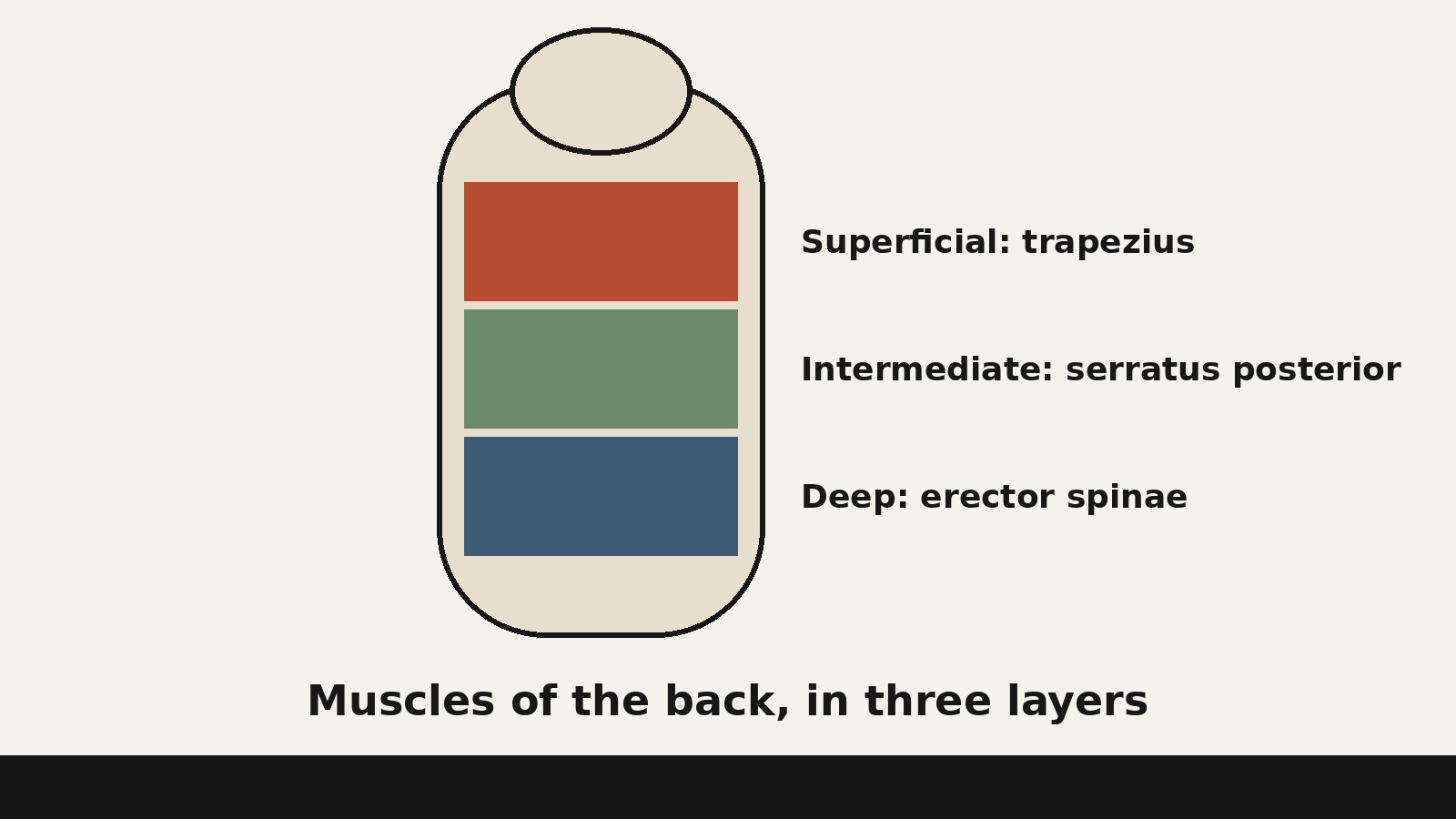
- The muscles of the back are the muscles that lie behind the vertebral column and around the shoulder blades, organized in three layers. The superficial layer moves the shoulder blades and arms and includes the trapezius, latissimus dorsi, levator scapulae, and rhomboid major and minor. The intermediate layer contains the serratus posterior superior and serratus posterior inferior, which act on the ribs. The deep layer, closest to the spine itself, includes the splenius muscles, the erector spinae, the transversospinales group, and small segmental muscles that link one vertebra to the next. Together these layers extend the trunk, bend it to the side, rotate it, move the shoulder blades, and help hold the trunk upright.
What Counts as a Muscle of the Back
The phrase “muscles of the back” is used in two senses. In the broad sense, it means the muscles you meet when you look at the back of the body, including large muscles that move the shoulder and arm. In the strict sense, only the intrinsic muscles count as true back muscles: the muscles that develop in the back itself and attach to the vertebral column. The superficial and intermediate muscles are grouped with the back because of where they lie and what they move, even though they develop elsewhere and are supplied by nerves from the front branches of spinal nerves. This article uses the broad, three layer system that most general anatomy sources use.
Muscle names often carry their region with them. Cervical means the neck, thoracic means the chest region of the spine, lumbar means the lower back, and sacral means the fused bones at the base of the spine. Longissimus thoracis therefore belongs to the chest region, while longissimus cervicis belongs to the neck. The word capitis means “of the head,” so a muscle ending in capitis reaches the skull. Four terms appear in every muscle description below. The origin is the attachment that moves least when the muscle contracts. The insertion is the attachment that moves most. The action is the movement the muscle produces. Innervation names the nerve that supplies the muscle. A muscle produces its action by pulling its insertion toward its origin.
Two more words need a plain definition. Unilateral means one side only. Bilateral means both sides together. Many back muscles rotate the head or bend the trunk to one side when they act alone, and extend the trunk or head, bending it backward, when they act together. That one rule explains a large share of back muscle behavior.
This article explains anatomy only and is not medical advice.
The Three Layers at a Glance
Anatomy organizes the muscles of the back by depth, from the surface inward. The superficial group is also called the appendicular group because its muscles are mainly associated with movement of the appendicular skeleton, the bones of the limbs and shoulder girdle, and because they are supplied by the anterior rami, the front branches, of spinal nerves, reflecting an origin outside the back itself. The intermediate group is also called the respiratory group because its two muscles act on the ribs and may serve a respiratory function, and it too is supplied by anterior rami. The deep group is also called the intrinsic group because its muscles originate in the back itself, and it is supplied by the posterior rami, the back branches, of spinal nerves.
| Layer | Muscles | Main job in plain language |
|---|---|---|
| Superficial | Trapezius, latissimus dorsi, levator scapulae, rhomboid major, rhomboid minor | Move and stabilize the shoulder blade; move the arm |
| Intermediate | Serratus posterior superior, serratus posterior inferior | Raise or lower the ribs |
| Deep, spinotransversales | Splenius capitis, splenius cervicis | Extend, bend, and rotate the head and neck |
| Deep, erector spinae | Iliocostalis, longissimus, spinalis | Extend and bend the trunk and neck; maintain the lumbar curve |
| Deep, transversospinales | Semispinalis, multifidus, rotatores | Extend, bend, and rotate the vertebral column; stabilize it |
| Deep, segmental | Interspinales, intertransversarii, levatores costarum | Link neighboring vertebrae; fine control of spinal position |
Depth is not a ranking of importance. Broad muscles produce large, visible movements of the shoulder and trunk. Deep muscles produce smaller, more precise adjustments and help keep the vertebral column steady while the larger muscles work.
The Superficial Layer: Muscles That Move the Shoulder
The superficial muscles are the first muscles under the skin of the back, and they are dominated by the shoulder blade, the scapula. The scapula is a flat, triangular bone that rests on the back of the rib cage, held in place almost entirely by muscle, since it has no bony joint with the rib cage. Its position therefore depends on the balance of the muscles that pull on it. In different combinations they raise the scapula, lower it, pull it toward the spine, move it away from the spine, or rotate it so the arm can be raised overhead.
Trapezius
The trapezius is the most superficial muscle of the upper back and one of the largest muscles in the body by area. Its name comes from its shape: the two trapezius muscles, left and right together, form a trapezoid, a four sided figure, across the upper back. Its origin includes the medial third of the superior nuchal line at the back of the skull, the external occipital protuberance, the ligamentum nuchae, and the spinous processes of the vertebrae from C7 to T12. Its insertion includes the lateral third of the clavicle, the medial side of the acromion, and the upper crest and tubercle of the spine of the scapula. The upper fibers elevate the scapula, the middle fibers retract it toward the midline, the lower fibers depress it, and together the fibers rotate it upward, the movement that lets the arm rise overhead. The trapezius also extends the head and neck. Its motor supply is the spinal accessory nerve, cranial nerve XI, with position sense fibers from cervical nerves C3 and C4, and it migrated during development from its level of origin in the neck to its final position, carrying its nerve and blood supply with it.
Latissimus Dorsi
The latissimus dorsi is the broad, flat muscle of the lower back, and it gives the back much of its width. Its origin includes the vertebral spines from T7 to the sacrum, the posterior third of the iliac crest, the top rim of the hip bone, the lower three or four ribs, and sometimes the inferior angle of the scapula. Its fibers converge into a tendon that inserts on the floor of the intertubercular groove of the humerus, the upper arm bone, and the tendon twists as it runs forward, so the fibers that originate highest end up inserting lowest. Despite lying on the back, the latissimus dorsi is mainly an arm muscle: it extends the arm backward, adducts it toward the body, rotates it medially, turning it inward, and depresses the shoulder. Anatomy teaching describes it as a prime mover of horizontal arm extension, the elbowing motion of pulling the elbow backward. It is supplied by the thoracodorsal nerve, from spinal levels C6 to C8 in most descriptions, and C7 and C8 in the university table used here.
Levator Scapulae
The levator scapulae is a smaller, strap like muscle at the side of the neck and upper back, and its name states its action: levator means lifter, and scapulae refers to the shoulder blade. It originates on the transverse processes, the side projections, of the first four cervical vertebrae, C1 to C4, and inserts on the medial border of the scapula from its superior angle down to the spine of the scapula. It elevates the scapula, and it also helps rotate the scapula and bend the neck to the same side. It is supplied by the dorsal scapular nerve, C5, with upper branches from C3 and C4. Because it connects the neck vertebrae directly to the shoulder blade, it links head and neck position to shoulder position.
Rhomboid Major and Rhomboid Minor
The rhomboids are two muscles that lie deep to the trapezius, between the spine and the scapula, and their name comes from their shape: a rhombus is a slanted, diamond like four sided figure. Rhomboid minor, the smaller, upper muscle, originates at the lower end of the ligamentum nuchae and the spinous processes of C7 and T1, and inserts on the medial border of the scapula at the root of its spine. Rhomboid major, the larger, lower muscle, originates on the spinous processes of T2 to T5, and inserts on the medial border of the scapula below the spine, down to the inferior angle. Both are supplied by the dorsal scapular nerve, C5, and both retract the scapula toward the spine, elevate it, and rotate it downward, the opposite rotation to the upward rotation that accompanies raising the arm. By pulling the scapula firmly against the rib cage and toward the midline, they give the shoulder a steady platform, and pulling the shoulder blades together is largely the rhomboids and the middle trapezius working together.
The Intermediate Layer: Two Muscles for the Ribs
Between the superficial muscles and the deep muscles lies a thin intermediate layer with only two muscles, the serratus posterior superior and the serratus posterior inferior. The word serratus means saw toothed, describing the way these muscles attach to the ribs in finger like slips. Both are supplied by ventral, front, branches of spinal nerves, not by the dorsal branches that supply the deep back muscles, and anatomy sources note that they are embryologically related to the intercostal muscles between the ribs rather than to the deep back muscles. That is why they are classed as intermediate rather than deep. The serratus posterior superior lies in the upper back, deep to the rhomboids: it originates on the ligamentum nuchae and the spinous processes of C7 and T1 to T3, inserts on the upper borders of ribs 1 to 4, elevates the upper ribs to enlarge the chest during inhalation, and is supplied by branches of the ventral primary rami of T1 to T4. The serratus posterior inferior lies at the junction of the chest and lower back: it originates on the thoracolumbar fascia and the spinous processes of T11 and T12 and L1 and L2, inserts on ribs 9 to 12, pulls the lower ribs downward to assist exhalation, and is supplied by branches of the ventral primary rami of T9 to T12.
The Deep Layer: Muscles That Move and Steady the Spine
The deep muscles are the intrinsic muscles of the back, the muscles that originate in the back and attach to the vertebral column itself. They are supplied by the posterior, dorsal, primary rami of the spinal nerves. OpenStax groups the muscles that move the vertebral column into five groups: the splenius group, the erector spinae with its three subgroups, the transversospinales, the segmental muscles, and the scalenes of the neck, which also contribute to deep inhalation. This article follows that framework for the back proper and treats the scalenes as neck muscles that overlap with it, rather than as back muscles in the strict sense. The deep muscles are also organized by fiber length. Longer, more superficial deep muscles span many vertebrae and produce larger movements. Shorter, deeper muscles span one or a few vertebrae and produce finer adjustments. That gradient is the key to the deep back: each length of muscle does a different part of the job of moving and steadying a column made of many separate bones.
Splenius: The Spinotransversales
The splenius muscles are the most superficial of the deep muscles, and they form the spinotransversales group. The name splenius means bandage, and it describes their broad, flat shape. Splenius capitis reaches the head: it originates on the ligamentum nuchae and the spinous processes of C7 to T6, and inserts on the mastoid process, the bony bump behind the ear, and the lateral end of the superior nuchal line of the skull. Splenius cervicis reaches the neck: it originates on the same region and inserts on the posterior tubercles of the transverse processes of C1 to C3. Both are supplied by the dorsal primary rami of spinal nerves C2 to C6, with splenius capitis receiving lateral branches of C2 and C3 in the StatPearls account. Acting on one side, each splenius muscle bends the head and neck to the same side and rotates the head to the same side. Acting on both sides together, the splenius muscles extend the head and neck, bending them backward. Splenius capitis lies deep to the trapezius, and splenius cervicis lies deep to the serratus posterior superior, the rhomboids, and the trapezius, so the splenius layer marks the boundary between muscles that move the shoulder and muscles that move the spine.
Erector Spinae: The Long Muscles of the Back
The erector spinae is the largest muscle group of the back by mass, and it is the primary extensor of the vertebral column. It forms the majority of the muscle mass of the back, it controls flexion, lateral flexion, and rotation of the vertebral column, and it maintains the lumbar curve, the inward curve of the lower back. Its origin includes the iliac crest, the sacrum, the transverse and spinous processes of vertebrae, and the supraspinal ligament along the tips of the spinous processes. Its insertion includes the angles of the ribs, the transverse and spinous processes of vertebrae, and the posterior aspect of the skull. It is supplied segmentally by the dorsal primary rami of spinal nerves from C1 to S5. Both sides together extend the trunk against gravity, one side alone bends the trunk to that side, and it also controls forward flexion by contracting in a controlled lengthening way as gravity pulls the trunk forward. The group is covered by the thoracolumbar fascia and by the serratus posterior inferior, the rhomboids, and the splenius muscles in its upper parts.
The erector spinae is three columns running side by side, from lateral to medial. Iliocostalis, the most lateral column, attaches to the ribs: it originates on the iliac crest and sacrum, inserts on the angles of the ribs, extends and laterally bends the trunk and neck, is supplied by the dorsal primary rami of C4 to S5, and is divided into lumborum, thoracis, and cervicis portions. Longissimus, the intermediate column and the longest of the three, originates on transverse processes at lower vertebral levels, inserts on transverse processes at higher levels and on the mastoid process, extends and laterally bends the trunk, neck, and head, is supplied by the dorsal primary rami of C1 to S1, and is divided into thoracis, cervicis, and capitis portions. Spinalis, the most medial column, originates on spinous processes at lower vertebral levels, inserts on spinous processes at higher levels and the base of the skull, extends and laterally bends the trunk and neck, is supplied by the dorsal primary rami of C2 to L3, and is divided into thoracis, cervicis, and capitis portions. Together the nine named parts in the OpenStax account cover the back from the sacrum and hip bone to the skull. Spinalis cervicis and capitis are the exception to that neat picture: they are poorly developed and not well defined, and they may be absent in some people.
Transversospinales: Short Muscles That Span a Few Vertebrae
Deep to the erector spinae lies the transversospinales group, named because its muscles run from the transverse process of one vertebra to the spinous process of a vertebra above. The group contains the semispinalis, the multifidus, and the rotatores, arranged from longest to shortest, and all three help extend, bend, and rotate the vertebral column and stabilize it. Semispinalis is the longest, spanning four to six segments: it originates on transverse processes of C7 to T12, inserts on the back of the skull for semispinalis capitis and on spinous processes four to six vertebrae above the origin for semispinalis cervicis and thoracis, extends and laterally bends the trunk, rotates the trunk to the opposite side, and is supplied by the dorsal primary rami of C1 to T12. Multifidus spans two to four segments: it originates on the sacrum and the transverse processes of C3 to L5, inserts on spinous processes two to four vertebral levels above its origin, extends and laterally bends the trunk and neck, rotates the trunk to the opposite side, and is supplied by the dorsal primary rami of C1 to L5, with the lumbar multifidus singled out in OpenStax for helping extend and laterally flex the vertebral column. Rotatores are the deepest and shortest, spanning one or two segments: they originate on transverse processes, insert on spinous processes one vertebra above for the short rotatores and two vertebrae above for the long rotatores, rotate the vertebral column to the opposite side, and are supplied by the dorsal primary rami of C1 to L5. Eleven pairs lie between T1 and T12 in the StatPearls account, and some pairs may be absent at the upper or lower end of the thoracic region, a normal variation.
Segmental Muscles: The Smallest Links
The deepest and smallest muscles of the back are the segmental muscles, which link one vertebra to the next. The interspinales run between the spinous processes of neighboring vertebrae: they originate on the upper border of a spinous process, insert on the lower border of the spinous process above, extend the trunk and neck, and are supplied by the dorsal primary rami of C1 to L5. The intertransversarii run between the transverse processes of neighboring vertebrae: they originate on the upper border of a transverse process, insert on the lower border of the transverse process above, laterally bend the trunk and neck, and are supplied by the same nerves. OpenStax groups them with the segmental muscles as important in stabilizing the vertebral column, bringing together the spinous and transverse processes of each consecutive vertebra. A third small group belongs here: the levatores costarum, the lifters of the ribs, which run from the transverse processes of thoracic vertebrae to the rib below. At the very top of the neck, the suboccipital muscles, including rectus capitis posterior major and minor and obliquus capitis superior and inferior, link the top two vertebrae to the skull and extend the head, with the major and obliquus muscles also rotating it to the same side, supplied by the suboccipital nerve, the dorsal ramus of C1. They are neck muscles in the strict sense, but they belong to the same design: short, deep muscles that control a small number of joints with precision.
How the Layers Work Together
No back movement uses one muscle alone. Extension of the trunk is produced mainly by the erector spinae acting on both sides, assisted by the semispinalis and multifidus and by the segmental muscles that keep each vertebra aligned while the larger muscles pull. Lateral flexion is produced by the muscles on the bending side acting together, with the superficial muscles setting the position of the shoulder blade. Rotation is produced by the transversospinales and the erector spinae acting in combination, with the multifidus and rotatores rotating the trunk to the opposite side and the splenius muscles rotating the head to the same side. Shoulder movement uses a different combination: elevation uses the trapezius upper fibers and the levator scapulae, retraction uses the rhomboids and the middle trapezius, upward rotation of the scapula uses the trapezius together with muscles of the chest wall, and downward rotation uses the rhomboids and the levator scapulae. Arm movement adds the latissimus dorsi, and breathing adds the intermediate layer, with the serratus posterior superior lifting the upper ribs for inhalation and the serratus posterior inferior drawing the lower ribs down for exhalation. In posture, the erector spinae maintains the lumbar curve, the transversospinales and segmental muscles keep each vertebra positioned relative to its neighbors, and the superficial muscles hold the shoulder blades in a balanced resting position, with the deep, short muscles making the finest adjustments and the long muscles making the larger ones.
Frequently Asked Questions
What are the three groups of back muscles?
The superficial group contains the trapezius, latissimus dorsi, levator scapulae, and rhomboid major and minor, and it mainly moves the shoulder blade and arm. The intermediate group contains the serratus posterior superior and inferior, and it acts on the ribs. The deep group contains the splenius muscles, the erector spinae, the transversospinales group, and the segmental muscles, and it moves and stabilizes the vertebral column. In the strict sense, only the deep, intrinsic muscles are considered true back muscles.
What is the largest muscle in the back?
What does the erector spinae do?
The erector spinae extends the vertebral column, bends it to the side, and rotates it, and it maintains the lumbar curve of the lower back. It has three columns: iliocostalis laterally, longissimus in the middle, and spinalis medially. Acting on both sides, it extends the trunk; acting on one side, it bends the trunk to that side; and it also controls forward bending as the trunk moves under gravity.
What is the difference between the trapezius and the latissimus dorsi?
The trapezius moves the shoulder blade: its upper fibers lift it, its middle fibers pull it toward the spine, its lower fibers pull it down, and together they rotate it upward. The latissimus dorsi moves the arm: it extends the arm backward, pulls it toward the body, and rotates it inward. The trapezius is supplied by the spinal accessory nerve, cranial nerve XI, while the latissimus dorsi is supplied by the thoracodorsal nerve.
What are the deep muscles of the back?
The deep muscles originate in the back and attach to the vertebral column, supplied by the posterior rami of spinal nerves. They include the splenius capitis and cervicis, the erector spinae with its iliocostalis, longissimus, and spinalis columns, the transversospinales group with semispinalis, multifidus, and rotatores, and the segmental muscles with interspinales and intertransversarii. Together they extend, bend, rotate, and stabilize the vertebral column.
Why do some back muscles have such long names?
Back muscle names combine the muscle name with its region, so a long name is usually a precise address. Longissimus thoracis means the longissimus muscle of the thoracic region, splenius capitis means the splenius muscle that reaches the head, and iliocostalis lumborum means the iliocostalis muscle of the lumbar region. The region words are cervical for the neck, thoracic for the chest region, lumbar for the lower back, sacral for the base of the spine, and capitis for the head.
Sources & references
- OpenStax, Anatomy and Physiology 2e, section 11.3, "Axial Muscles of the Head, Neck, and Back," (axial versus appendicular organization; splenius group; erector spinae as primary extensor of the vertebral column, controlling flexion, lateral flexion, and rotation, and maintaining the lumbar curve; iliocostalis, longissimus, and spinalis columns and their regional parts; transversospinales running from transverse to spinous processes; semispinalis parts; lumbar multifidus; segmental muscles bringing together spinous and transverse processes of consecutive vertebrae; scalenes contributing to deep inhalation)
- OpenStax, Anatomy and Physiology 2e, Chapter 11 chapter review, (five groups of muscles that move the vertebral column: splenius, erector spinae subgroups, transversospinales, segmental muscles, and scalenes, with regional parts listed)
- University of Arkansas for Medical Sciences, "Muscles of the Back Region" and "Muscles of the Upper Limb That Are Dissected in the Back Region," (origins, insertions, actions, innervation, and notes for trapezius, latissimus dorsi, levator scapulae, rhomboid major and minor, serratus posterior superior and inferior, erector spinae and its columns, interspinales, intertransversarii, multifidus, rotatores, semispinalis, spinalis, splenius capitis and cervicis, suboccipital muscles, and serratus anterior as an upper limb muscle dissected in the back region)
- Henson, Kadiyala, and Edens, "Anatomy, Back, Muscles," StatPearls, (three group introduction and the strict sense in which only intrinsic muscles are true back muscles; dorsal rami supplying intrinsic muscles and ventral rami supplying trapezius, latissimus dorsi, levator scapulae, and rhomboids; splenius capitis and cervicis origins, insertions, and unilateral versus bilateral actions; erector spinae regional groups; spinalis thoracis details; longissimus and iliocostalis regional parts; transversospinales lengths, with rotatores spanning one to two segments, multifidus two to four, and semispinalis four to six; variants including absent or variable splenius attachments and absent rotatores pairs)
- Chabot College, Medicine LibreTexts, "Muscles of the Axial Skeleton," (latissimus dorsi as a broad, flat muscle of the lower back and prime mover of horizontal arm extension; trapezius targeting the spine and shoulders; erector spinae as the majority of back muscle mass and primary extensor, with iliocostalis lateral, longissimus intermediate, and spinalis medial)


