Short answer
- The function of the breathing system is to move oxygen into the body and carbon dioxide out of it. Air travels through the nose or mouth, down the windpipe, and into the lungs, where oxygen passes into the blood at tiny air sacs called alveoli and carbon dioxide passes out of the blood to be breathed out. The diaphragm and the muscles between the ribs power this movement, and control centers in the brainstem adjust the rate and depth of breathing to match the body's changing needs. The system also warms, moistens, and filters incoming air, helps produce speech, supports the sense of smell, and helps keep the acidity of the blood in balance. # What Is the Function of the Breathing System? Every breath does one central job, in partnership with the rest of the body. The breathing system, also called the respiratory system, brings air into the lungs, swaps gases between that air and the blood, and sends used air back out. It works hand in hand with the circulatory system, which carries the gases to and from every tissue, and with the brain, which sets the rhythm and adjusts it minute by minute. This article follows a breath from the nose or mouth to the alveoli, shows how gases cross into and out of the blood, describes the muscles and pressure changes that move air, and explains how the brain keeps the process matched to the body's needs. It also covers the system's secondary jobs and common misunderstandings.
The One Job, in Four Steps
Physiology texts break respiration into four linked processes, each in a different place and depending on the step before it.
| Step | What happens | Where it happens |
|---|---|---|
| 1. Pulmonary ventilation | Air moves into and out of the lungs | Airways, lungs, and breathing muscles |
| 2. External respiration | Oxygen and carbon dioxide are exchanged between the lungs and the blood | Alveoli and the capillaries around them |
| 3. Gas transport | Gases travel through the bloodstream | Blood vessels, carried by the blood |
| 4. Internal respiration | Oxygen and carbon dioxide are exchanged between the blood and the tissues | Capillaries in the tissues throughout the body |
Pulmonary ventilation is what people usually mean when they say breathing: the mechanical part. External respiration is the chemical exchange at the lungs. Gas transport is the delivery service. Internal respiration is the matching exchange at the far end, where cells receive oxygen and release carbon dioxide. If any one step stalls, the others cannot complete the job alone.
Two similar words are worth keeping apart. Breathing is the movement of air. Cellular respiration is the set of chemical reactions inside cells that uses oxygen to release energy and produces carbon dioxide as a waste product. The breathing system serves cellular respiration but does not perform it: the lungs supply the oxygen and remove the carbon dioxide, and the cells do the chemistry.
The Air Pathway: From Nose and Mouth to Alveoli
Air enters through the nose or the mouth. The nose is the usual entry point at rest. The National Heart, Lung, and Blood Institute (NHLBI) notes that the nose and mouth moisten and warm the air, because cold, dry air can irritate the lungs. The mouth takes over when more air is needed, for example during vigorous activity, but does less warming and filtering.
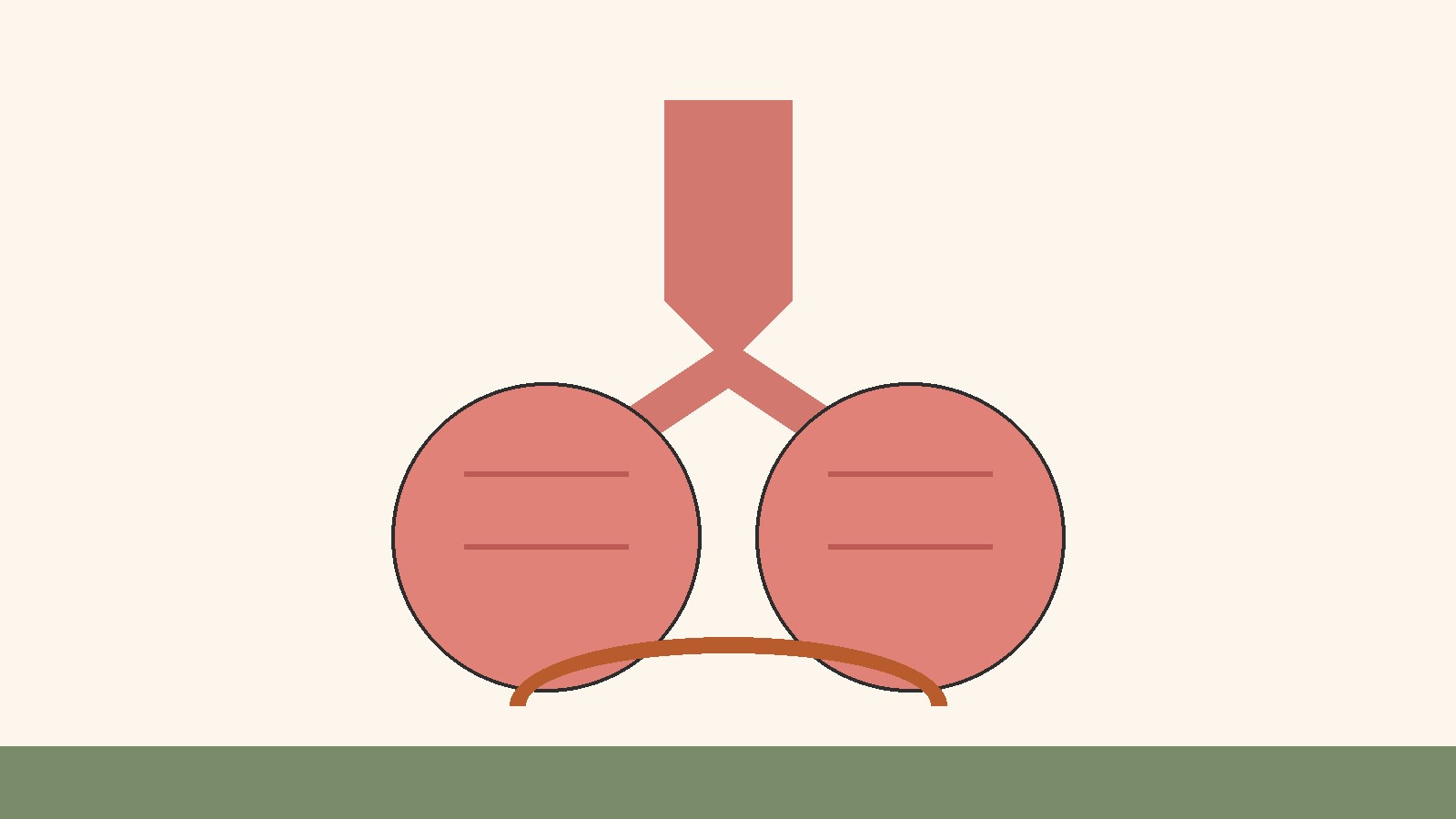
From the nose or mouth, air passes through the pharynx, the passage at the back of the throat, then past the larynx, or voice box, into the trachea, the windpipe. The trachea is held open by rings of cartilage, and at its lower end it divides into two bronchi, one per lung. Cleveland Clinic calls this branching network the tracheobronchial tree: increasingly smaller tubes carrying air toward the small air sacs in the lungs.
Inside each lung, the bronchi divide into narrower branches called bronchioles, ending in clusters of tiny air sacs called alveoli. The NHLBI puts the number of alveoli at about 150 million and describes them as elastic. Their insides are coated with surfactant, which helps the lungs inflate more easily on inhalation and helps prevent collapse on exhalation. The branching design spreads inhaled air across an enormous surface area in the limited space of the chest, so gas exchange can happen quickly and on a large scale.
Not every part of the pathway exchanges gas. The nose, pharynx, larynx, trachea, bronchi, and larger bronchioles form the conducting zone, which delivers air to the alveoli warmed, moistened, and cleaned. Only the respiratory zone, the smallest bronchioles, the alveolar ducts, and the alveoli themselves, takes part in gas exchange. Air in the conducting airways never meets the blood, so it is sometimes called dead space air. That is normal anatomy: the conducting zone protects the delicate exchange surfaces by doing the conditioning and filtering first.
The lungs are the main organs, but not the whole system. Cleveland Clinic lists its parts as the nose and nasal cavity, the mouth and oral cavity, the sinuses, the pharynx, the larynx, the trachea, the bronchi, the lungs, and the diaphragm. The ribs and chest wall protect the lungs and give the breathing muscles something to pull against, and the blood vessels, nerves, and muscles of breathing are part of the working system too, even though they are not airways.
Gas Exchange: How Oxygen Gets In and Carbon Dioxide Gets Out
Gas exchange is the heart of the breathing system’s function, and it happens by diffusion: the movement of molecules from an area of higher concentration to an area of lower concentration. No pump pushes oxygen across the alveolar wall, and no pump pushes carbon dioxide back. The gases move on their own, down their concentration gradients, across membranes only a few cells thick.
Each alveolus is wrapped in capillaries, tiny blood vessels. The NHLBI explains the sequence: the pulmonary artery delivers blood rich in carbon dioxide and low in oxygen to the capillaries around the alveoli. Oxygen then moves from the alveoli into the blood, while carbon dioxide moves from the blood into the alveoli. The refreshed blood returns toward the heart, which pumps it to the rest of the body, and the carbon dioxide rich air is breathed out on the next exhalation.
Three features make this exchange efficient: the alveolar and capillary walls are extremely thin, so gases cross a short distance; the total surface area is very large, because there are so many alveoli; and blood flows past continuously, so refreshed blood is carried away and replaced by blood that still needs oxygen, keeping the concentration gradients pointing the right way.
The lungs do not create oxygen or destroy carbon dioxide. They collect oxygen from the air and hand it to the blood, and collect carbon dioxide from the blood and release it into exhaled air. The system is a gateway and an exchange point, not a factory.
Breathing Mechanics: The Muscles and Pressure That Move Air
The lungs themselves do not move air. They are passive, elastic structures that follow the movements of the chest wall. Air moves because the volume of the chest cavity changes, and when volume changes, pressure changes with it. Air always flows from higher pressure to lower pressure, so changing the pressure inside the lungs relative to the air outside is enough to drive air in and out.
Inhalation, also called inspiration, begins when the diaphragm contracts. The diaphragm is a large, dome shaped muscle that forms the floor of the chest cavity. When it contracts, it flattens and moves downward, which enlarges the chest cavity. At the same time, the external intercostal muscles, the muscles between the ribs, contract and move the ribs upward and outward, expanding the rib cage further. Because the lungs are coupled to the chest wall by the pleural fluid in the space around them, the expanding chest pulls the lungs open with it. As lung volume increases, pressure inside the alveoli falls below atmospheric pressure, and air flows in to equalize the difference.
Exhalation, also called expiration, is normally passive at rest. When the diaphragm and intercostal muscles relax, the elastic tissue of the lungs recoils and the chest cavity shrinks. Lung volume falls, pressure inside the alveoli rises above atmospheric pressure, and air flows out. No muscle effort is needed to push quiet exhalation along. The stored elastic energy of the stretched lungs and chest wall does the work.
Forced breathing is different: neck muscles help lift the chest wall further during forced inhalation, while abdominal muscles push upward against the diaphragm and internal intercostal muscles compress the rib cage during forced exhalation. Forced breathing uses muscle power in both directions; quiet breathing uses it mainly for inhalation.
The relationship between volume and pressure follows Boyle’s law, which states that at a constant temperature, the pressure of a gas is inversely related to its volume: when volume increases, pressure decreases, and when volume decreases, pressure increases. OpenStax, the Rice University based open textbook program, uses this principle to explain pulmonary ventilation in full: inspiration lowers pressure inside the alveoli below atmospheric pressure so air flows in, and expiration raises it above atmospheric pressure so air flows out. The muscles change the size of the container, and pressure does the rest.
Surfactant solves a mechanical problem: the inner surface of each alveolus is wet, and surface tension tends to pull small air sacs closed. Surfactant, secreted by specialized cells in the alveolar lining, lowers that surface tension. Without it, the alveoli would be far more likely to collapse during exhalation, and each inhalation would require much more effort.
How Much Air Moves: Volumes and Rates
A normal quiet breath moves a modest amount of air. OpenStax gives tidal volume, the amount of air that normally enters the lungs during quiet breathing, as about 500 milliliters. Deeper breaths move more: inspiratory reserve volume is the extra air that can be drawn in past a normal inhalation, and expiratory reserve volume is the extra air that can be forced out past a normal exhalation. Residual volume is the air that remains in the lungs even after the fullest possible exhalation. It never leaves, and it serves a purpose: it keeps the alveoli from collapsing completely between breaths.
Total lung capacity, the total amount of air the lungs can hold after a forceful inhalation, is about 6,000 milliliters for males and about 4,200 milliliters for females in the OpenStax figures. Vital capacity, the amount of air that can be moved into or out of the lungs, is between 4,000 and 5,000 milliliters. These are population figures, not personal targets, since lung size varies with body size, age, sex, and other factors. They show the scale of the system: a resting breath uses only a small fraction of what the lungs can hold, leaving a large reserve.
Breathing rate is the number of complete breathing cycles per minute. Cleveland Clinic’s published table of typical breathing rates gives 12 to 20 breaths per minute for adults aged 18 to 50, and 13 to 20 breaths per minute for adults aged 50 and up. OpenStax gives a similar adult range of 12 to 18 breaths per minute, noting that the rate is higher in children and falls toward the adult range through adolescence. Small differences between published ranges are normal. At rest, an adult breathes roughly once every three to five seconds, and that pace is set automatically.
Rate and depth work together. The total air moved per minute, called minute ventilation, is the breathing rate multiplied by the volume of each breath. The body can raise minute ventilation by breathing faster, by breathing deeper, or by doing both. During physical activity, both usually rise, because working muscles consume more oxygen and produce more carbon dioxide.
Working With Circulation: The Partnership That Completes the Job
The breathing system cannot finish its job alone, and this is the part of the story that is easiest to miss. Oxygen that reaches the blood in the lungs still has to reach the tissues, and carbon dioxide produced in the tissues still has to reach the lungs. The circulatory system does that carrying, and the two systems are arranged so that their flows meet at exactly the right places.
The route runs in a loop. Blood low in oxygen and rich in carbon dioxide returns to the right side of the heart, which pumps it through the pulmonary artery to the lungs. In the capillaries around the alveoli, gas exchange refreshes it. The refreshed blood travels through the pulmonary veins to the left side of the heart, which pumps it through the aorta and arteries to the tissues. In the tissue capillaries, the exchange runs in reverse, and the blood then returns to the right side of the heart.
Inside the blood, gases travel in different forms. Most oxygen binds to hemoglobin, the iron containing protein inside red blood cells. Each hemoglobin molecule can carry up to four oxygen molecules, and the binding is reversible: hemoglobin picks up oxygen where oxygen is plentiful, in the lungs, and releases it where oxygen is scarcer, in the tissues. A small amount of oxygen also travels dissolved directly in the plasma, the liquid part of the blood, but hemoglobin carries the large majority. For the return journey, the National Center for Biotechnology Information’s StatPearls summary of carbon dioxide transport describes three forms: dissolved in plasma, bound to hemoglobin, and, in the largest share, converted to bicarbonate ions inside red blood cells with the help of the enzyme carbonic anhydrase. At the lungs, the bicarbonate reaction reverses, recreating carbon dioxide, which diffuses into the alveoli and is breathed out.
Breathing rate alone does not determine how much oxygen the tissues receive. Heart rate, blood flow, and hemoglobin levels matter too. The breathing system sets the gas composition of the blood leaving the lungs; the circulatory system decides how quickly that blood reaches each tissue. When demand rises, both respond together.
Control of Breathing: How the Brain Sets the Rhythm
Breathing usually happens without thought. That automatic rhythm comes from respiratory centers in the brainstem, the lower part of the brain that connects to the spinal cord. OpenStax identifies two main regions: the medulla oblongata and the pontine respiratory group in the pons. The medullary respiratory center sets the basic rhythm. Its dorsal respiratory group maintains a constant rhythm by stimulating the diaphragm and intercostal muscles to contract for inspiration; when its activity pauses, those muscles relax and expiration follows. Its ventral respiratory group becomes more active during forced breathing, stimulating the accessory muscles used for deeper inhalation and forceful exhalation. The pontine respiratory group modifies the medullary rhythm: its pneumotaxic center limits inspiration and helps set the overall rate, while its apneustic center supports deeper inspiration.
The strongest adjusting signal is not oxygen, but carbon dioxide. OpenStax states that the major factor stimulating the medulla and pons is blood carbon dioxide concentration, not oxygen. Chemoreceptors detect changes in carbon dioxide, hydrogen ions, and pH. Central chemoreceptors in the brain and brainstem respond mainly to hydrogen ion levels, which rise when carbon dioxide rises; peripheral chemoreceptors in the carotid arteries and aortic arch monitor the blood directly. When carbon dioxide rises, breathing quickens and deepens, blowing off more carbon dioxide and bringing its level back down. When it falls, breathing slows and shallows. It is a negative feedback loop.
Oxygen plays mainly a backup role. OpenStax notes that peripheral chemoreceptors stimulate increased breathing when blood oxygen falls to about 60 millimeters of mercury or less, a large drop. Because most oxygen travels bound to hemoglobin and chemoreceptors sense only dissolved oxygen, a substantial fall is needed first. In everyday conditions, carbon dioxide does the fine tuning.
Automatic control is not inflexible. Higher brain regions can override the brainstem rhythm briefly, which is why you can hold your breath, speak, sing, or cough on purpose. The hypothalamus and related regions also adjust breathing for emotion, pain, and body temperature, and stretch receptors in the lungs help prevent overinflation. It is a layered system: an automatic rhythm generator, chemical sensors that tune it continuously, and higher centers that can take over briefly.
The Secondary Jobs: Conditioning, Protection, Speech, and Smell
Gas exchange is the main function, but the breathing system does several other jobs at the same time, most of them along the air pathway before air reaches the alveoli.
The first is conditioning the air. The nose, sinuses, and upper airways warm inhaled air toward body temperature and add moisture to it, and Cleveland Clinic adds that the sinuses help regulate its temperature and humidity. Warm, moist air is gentler on the delicate alveolar surfaces and keeps airway mucus at the right consistency for filtering. Mouth breathing bypasses much of this conditioning, which is one reason it feels harsher in cold air.
The second is protection. The airways are lined with mucus and cilia, tiny hairlike structures that beat in coordinated waves. Cleveland Clinic describes the arrangement: cilia filter out dust, germs, and other irritants, and material that does get in is trapped in mucus and moved outward to be expelled by coughing or sneezing. Those reflexes are part of the defenses, not failures of them.
The third is speech. Air moving out of the lungs passes through the larynx, where it vibrates the vocal cords and produces sound. The shape of the throat, mouth, and nasal passages then modifies that sound into speech. Without controlled exhalation, sustained speech would be impossible: speakers unconsciously adjust the depth and timing of their breaths to fit sentences, pausing to inhale at natural breaks.
The fourth is smell. When air is drawn through the nose, odor molecules pass over the olfactory receptors high in the nasal cavity, which send signals to the brain about what is being smelled. Smell and taste work together closely, which is why food seems flavorless when the nose is blocked. The breathing system’s role here is simple but essential: it delivers the odor molecules to the receptors.
The fifth is helping to balance blood acidity. Cleveland Clinic lists this among the system’s functions: too much carbon dioxide lowers the blood’s pH, making it more acidic, and removing carbon dioxide helps maintain acid base balance. The kidneys handle the longer term side of this balance; breathing provides the fast adjustment.
Breathing at Rest and During Activity
At rest, the system runs quietly and efficiently. Quiet breathing, called eupnea in physiology texts, uses the diaphragm and external intercostal muscles for inhalation and elastic recoil for exhalation. The rate sits in the adult resting range described earlier, roughly 12 to 20 breaths per minute, and each breath moves about 500 milliliters of air. Most of the lung volume is unused at rest, leaving a large reserve.
When the body becomes more active, demand changes on both sides of the exchange. Working muscles consume more oxygen and produce more carbon dioxide. Carbon dioxide rises in the blood, the chemoreceptors detect it, and the respiratory centers increase both the rate and the depth of breathing. The heart responds in parallel, pumping more blood through the lungs and tissues. The adjustment is graded: light activity produces a modest increase, and heavier activity produces a larger one, up to the point where forced breathing brings in the accessory muscles.
Common Misunderstandings About the Breathing System
“Breathing in more air means more oxygen for the body.” Not exactly. What matters is how much oxygen reaches the blood and then the tissues, which depends on gas exchange, blood flow, and hemoglobin, not on the raw volume of air moved. Very rapid, shallow breathing can move a lot of air through the conducting airways without adding much to alveolar ventilation, because much of each small breath stays in the dead space. Depth matters as much as rate.
“The lungs pump air like a bellows.” The lungs do not generate the movement. The diaphragm and chest muscles change the size of the chest cavity, and pressure differences move the air. The lungs follow passively, pulled open by the chest wall and recoiling by their own elasticity.
“We breathe only to get oxygen.” Oxygen intake is half the job. Removing carbon dioxide is equally important, because carbon dioxide accumulation acidifies the blood and disrupts the chemical environment cells need. The control system treats carbon dioxide as its main signal precisely because its removal matters so much.
“Holding your breath starves the body of oxygen first.” The urgent urge to breathe during a held breath is driven mainly by rising carbon dioxide, not by falling oxygen. Oxygen falls too, but the carbon dioxide signal arrives first and strongest in most situations.
“Breathing is entirely automatic.” Breathing is mainly automatic, but also partly voluntary. You can hold your breath, speak, sing, and cough on purpose, using the same muscles and airways. The automatic system resumes control when the voluntary override ends, because the chemical drive to breathe cannot be ignored indefinitely.
Frequently Asked Questions
What is the main function of the breathing system?
Its main function is gas exchange: bringing oxygen into the body and removing carbon dioxide. Oxygen passes from the alveoli into the blood, carbon dioxide passes from the blood into the alveoli, and the carbon dioxide is breathed out. The system also warms, moistens, and filters incoming air, helps produce speech, supports smell, and helps balance blood acidity.
What is the difference between breathing and respiration?
Breathing, or pulmonary ventilation, is the mechanical movement of air into and out of the lungs. Respiration is broader: ventilation, gas exchange at the lungs, gas transport in the blood, gas exchange at the tissues, plus the cellular reactions that use oxygen and produce carbon dioxide. Breathing is one step within respiration.
How does gas exchange work in the alveoli?
Gas exchange works by diffusion across very thin membranes. Oxygen is more concentrated in the alveoli than in arriving blood, so it diffuses into the capillaries. Carbon dioxide is more concentrated in that blood than in the alveoli, so it diffuses the other way. The many alveoli provide a huge surface area, and continuous blood flow keeps the gradients pointing in the right direction.
What does the diaphragm do when you breathe in?
When the diaphragm contracts, it flattens and moves downward, enlarging the chest cavity. The external intercostal muscles expand the rib cage at the same time. The larger chest volume lowers the pressure inside the lungs below atmospheric pressure, and air flows in. When the diaphragm relaxes, the chest cavity shrinks, pressure rises, and air flows out.
What controls how fast you breathe?
Respiratory centers in the brainstem, mainly the medulla oblongata and the pons, set the basic rhythm. Chemoreceptors in the brain, the carotid arteries, and the aortic arch monitor carbon dioxide, hydrogen ions, pH, and oxygen in the blood. Rising carbon dioxide is the strongest signal to breathe faster and deeper. Higher brain regions can override the rhythm briefly for speech, singing, or holding the breath, and emotion and body temperature also influence the rate.
How does oxygen get from the lungs to the body’s tissues?
Oxygen passes from the alveoli into the surrounding capillaries, where most of it binds to hemoglobin inside red blood cells. The blood travels through the pulmonary veins to the left side of the heart, which pumps it through the arteries to the tissues, where hemoglobin releases the oxygen. Carbon dioxide follows the reverse route back to the lungs.
Why does breathing get faster during exercise?
Working muscles use more oxygen and produce more carbon dioxide. Rising carbon dioxide is detected by chemoreceptors, which signal the brainstem centers to increase the rate and depth of breathing, while the heart increases its output. The adjustment is automatic and graded to the level of activity.
Does the breathing system do anything besides gas exchange?
Yes. It warms and moistens inhaled air, filters particles with mucus and cilia, produces the airflow behind speech, delivers odor molecules to the smell receptors, and helps maintain acid base balance by controlling carbon dioxide.
Sources & references
- National Heart, Lung, and Blood Institute (NHLBI), National Institutes of Health, "How the Lungs Work: The Respiratory System." Opened and read October 25, 2026. Airways carry oxygen rich air to the alveoli and carbon dioxide out of the lungs; air enters through the nose or mouth, which moistens and warms it; bronchial tubes branch into bronchioles ending in alveoli; about 150 million alveoli; surfactant reduces the work of breathing and prevents collapse on exhalation; pulmonary artery blood is rich in carbon dioxide and low in oxygen; oxygen moves from alveoli into blood while carbon dioxide moves from blood into alveoli.
- Cleveland Clinic, "Respiratory System: Organs, Facts, Anatomy and Function." Opened and read October 25, 2026. Main function is gas exchange, bringing oxygen into the lungs and moving carbon dioxide out, working closely with the circulatory system; diaphragm pulls down to create space in the chest and draw air in; air travels through the mouth or nose, trachea, bronchi, and bronchioles to the alveoli; carbon dioxide returns through the bloodstream and leaves as the diaphragm moves back up; cilia filter dust, germs, and irritants, and mucus traps material that cilia move out by coughing or sneezing; sinuses help regulate temperature and humidity of inhaled air; too much carbon dioxide lowers blood pH, and removing it helps maintain acid base balance.
- Cleveland Clinic, "Bradypnea." Used for the typical breathing rate table only, via search result summary (page not opened in full in this pass). Typical rates: ages 18 to 50, 12 to 20 breaths per minute; ages 50 and up, 13 to 20 breaths per minute.
- OpenStax, Anatomy and Physiology 2e, section 22.4, "The Process of Breathing." Opened and read October 25, 2026. Pulmonary ventilation driven by pressure differences; Boyle's law, pressure inversely related to volume; diaphragm and external intercostals drive normal inspiration, expiration normally passive by elastic recoil, accessory muscles used in forced breathing; surfactant from type II alveolar cells reduces surface tension and prevents alveolar collapse; tidal volume about 500 milliliters; total lung capacity about 6,000 milliliters for males and about 4,200 milliliters for females; vital capacity between 4,000 and 5,000 milliliters; respiratory center in the medulla oblongata responds primarily to carbon dioxide, oxygen, and pH; medullary dorsal and ventral groups and pontine pneumotaxic and apneustic centers; carbon dioxide is the major stimulus, oxygen stimulates mainly when it falls to about 60 millimeters of mercury or less; hypothalamus and limbic regions adjust breathing for emotion, pain, and temperature.
- National Center for Biotechnology Information, StatPearls, "Physiology, Carbon Dioxide Transport." Opened and read October 25, 2026, via search result summary. Carbon dioxide is transported dissolved in plasma, bound to hemoglobin, and, in the largest share, as bicarbonate formed inside red blood cells by carbonic anhydrase; at the lungs the reaction reverses and carbon dioxide diffuses into the alveoli to be exhaled.
- OpenStax, Anatomy and Physiology 2e, transport of gases material, via search result summary. Oxygen is transported primarily bound to hemoglobin in red blood cells, each hemoglobin molecule carrying up to four oxygen molecules; carbon dioxide returns dissolved in plasma, bound to hemoglobin, or mostly converted to bicarbonate.


