Short answer
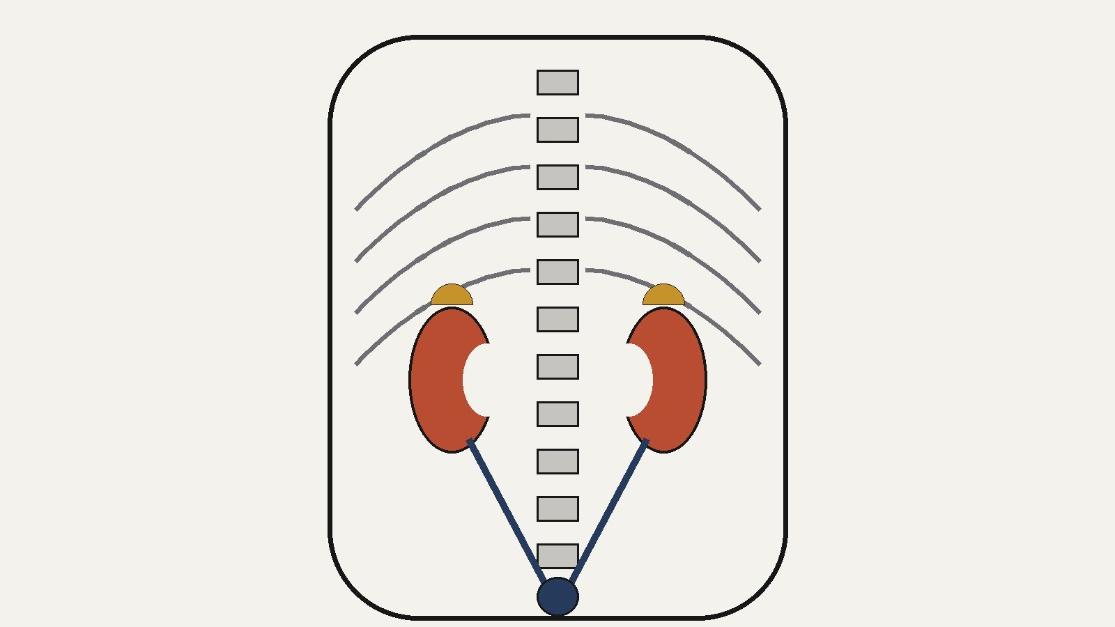
- Your kidneys sit in the upper abdomen, but toward the back, not the front. Each one lies just below the rib cage, on either side of your spine, in the area of the lower back. The right kidney usually sits a little lower than the left because the liver takes up space above it. Both kidneys rest against the back wall of the abdomen, behind the membrane that lines the abdominal cavity, which is why they are called retroperitoneal organs. This article describes general anatomy only; if you have questions about your health, talk to a qualified health professional.
Ask where the kidneys are and most people gesture vaguely toward their lower back. That is closer than many anatomical guesses get, but the details are more interesting than the gesture. The kidneys are not in the abdomen in the way the stomach or intestines are in the abdomen. They sit behind that scene, tucked against the back wall, protected by the lowest ribs and a cushion of fat, each one anchored in a precise position that explains why they are where they are. This article maps that position: the exact region of the body they occupy, the difference between the left and the right, the layered wrapping that holds each one in place, the organs that neighbor them, and, at a general level, what they do once situated there.
The Simple Answer: Below the Ribs, Beside the Spine
The National Institute of Diabetes and Digestive and Kidney Diseases, part of the National Institutes of Health, describes the kidneys as two bean-shaped organs, each about the size of a fist, located just below the rib cage, one on each side of the spine. That sentence alone answers the most common version of the question, and it is worth unpacking each clause.
“Just below the rib cage” sets the vertical level. The kidneys occupy the upper part of the abdomen, roughly at the level of the waist or just above it. “One on each side of the spine” sets the horizontal position: left and right, hugging the midline without touching it. And although the kidneys are abdominal organs, they are not floating freely among the other abdominal organs. They sit at the back of the space, against the posterior abdominal wall, deep to the muscles and fat of the lower back. This is why the kidneys are often described as lower-back organs: from the outside, the nearest surface of the body is the back, not the belly.
Anatomy textbooks pin the position more precisely using the spine as a ruler. The left kidney lies at about the level of the twelfth thoracic vertebra to the third lumbar vertebra, abbreviated T12 to L3, and the right kidney sits slightly lower. The upper portions of both kidneys are partially protected by the eleventh and twelfth ribs, the two shortest ribs, which float freely at the bottom of the rib cage. So when you feel the lowest ribs on your back, your kidneys are just beneath them, on either side of the spine.
Why the Kidneys Are Called Retroperitoneal
Here is the detail that separates the kidneys from most of the organs people picture when they think of the abdomen. The abdominal cavity is lined by a smooth membrane called the peritoneum, and organs such as the stomach, liver, and much of the intestine sit inside that lining, wrapped by it. The kidneys do not. They lie behind the peritoneum, between the parietal peritoneum (the part of the membrane that lines the back wall) and the muscles of the posterior abdominal wall. That position has a one-word name: retroperitoneal, literally “behind the peritoneum.”
The distinction matters for understanding the layout of the body. The kidneys share the retroperitoneal space with a few other structures that run along the back wall, including the abdominal aorta and the inferior vena cava, the great vessels that carry blood to and from the lower body. The ureters, the tubes that carry urine from the kidneys to the bladder, are also retroperitoneal. The bladder itself sits lower, in the pelvis, with only its dome covered by peritoneum. So the urinary system is mostly a behind-the-scenes affair: the filters and the pipes are retroperitoneal, and only the storage bag peeks into the main abdominal cavity.
For a lay reader, “retroperitoneal” can be read as “deep and toward the back.” It is one reason the kidneys are better protected than their job might suggest. They are not exposed to the jostling of the intestines, and they sit in a recess cushioned by fat and muscle.
Left and Right: Almost Symmetrical, But Not Quite
The two kidneys are a matched pair in the way your hands are a matched pair: the same design, mirrored, with a small asymmetry forced by the neighbors. The right kidney usually sits slightly lower than the left. The cause is the liver. The liver is a large organ that occupies the upper right of the abdomen, and it simply crowds the space above the right kidney, pushing it downward a little. OpenStax Anatomy and Physiology describes the right kidney as lower “due to slight displacement by the liver.”
How much lower? Sources vary, and the variation itself is the honest answer. The displacement is usually described as roughly one vertebra’s worth of height, though it differs between individuals. It is a tendency, not a rule: imaging studies and dissections show that the exact heights of the two kidneys vary from person to person, and a slightly larger difference in one individual does not by itself mean anything about either kidney.
The asymmetry has a couple of practical consequences worth noting for anatomy students. Because the liver bulges downward on the right, the right kidney is tucked a bit more deeply under the rib cage, while the left kidney, sitting slightly higher, has its upper end near the stomach, spleen, and pancreas on the left side. The two kidneys therefore face slightly different neighbors, which is covered in the section below. And because the right kidney sits lower, the right ureter is typically a touch shorter than the left, though both are about the same length overall.
The Position Is Set Before Birth
The kidneys do not start out where you find them in an adult. Early in fetal development, the permanent kidneys form low down, in the pelvic region, and then ascend to their final position in the lumbar region as the body grows. The ascent happens at roughly weeks six to nine of gestation: the kidneys rise from their initial pelvic location to the upper retroperitoneum, while the body lengthens around them. Part of the climb is genuine movement, and part is the surrounding body growing taller and carrying the landmarks upward, which is why anatomy teaching often describes the kidneys as being carried up by the growth of the lumbar and sacral regions.
The ascent explains the plumbing as well. As each kidney rises, it draws its blood supply from the vessels nearest to it at each stage, and by the time it reaches the lumbar region, the definitive supply arrives from the aorta through the renal arteries. The kidney also rotates during the ascent, turning so that the renal pelvis ends up facing medially, toward the midline, which is the orientation seen in the adult hilum.
Occasionally the ascent stops early and a kidney remains lower than usual, in the pelvis or lower abdomen. That is an anatomical variation, not something a reader can feel or change, but it is one more reason the exact position of the kidneys can differ from person to person. The layout described in this article is the usual endpoint of that early climb.
The Layers That Hold Each Kidney in Place
A kidney does not just rest where it is; it is wrapped and anchored. Three layers surround each kidney, listed from the organ outward:
- The renal capsule, a tough, thin coat of dense fibrous connective tissue that directly covers the kidney. It holds the organ’s shape and protects the soft tissue inside.
- The renal fat pad (also called perirenal fat), a shock-absorbing layer of adipose tissue around the capsule. It cushions the kidney against bumps and movement.
- The renal fascia, a tough outer layer of connective tissue that anchors the whole package. The fascia, together with the overlying peritoneum, fastens the kidney firmly to the posterior abdominal wall in its retroperitoneal position.
Beyond these layers, the lower ribs guard the upper portions of the kidneys, and the muscles and fat of the back provide the rest of the protection. OpenStax summarizes the arrangement by saying the kidneys are “well protected by muscle, fat, and ribs.” The kidneys can shift slightly when you breathe or change posture, since they are cushioned rather than bolted down, but the fascia and surrounding tissue keep them in their region.
The Neighborhood: What Sits Near the Kidneys
The kidneys occupy a crowded part of the body, and the neighbors differ on the two sides. The single most constant neighbor is the adrenal gland: one sits on the upper end of each kidney, as the name suggests (adrenal means “on top of renal”). The adrenal glands are separate organs with their own jobs in hormone production, but anatomically they cap each kidney, and the whole kidney-plus-gland unit shares the surrounding fat and fascia.
The rest of the neighborhood, as described in standard anatomy references, runs roughly as follows. On the right side, the kidney lies behind the liver, which sits above and in front of it; the duodenum, the first part of the small intestine, and the ascending colon, the first part of the large intestine, are also nearby on the right. On the left side, the kidney sits behind and below the spleen and the stomach, with the pancreas and the descending colon close by on the left. Running down the middle, between the two kidneys, are the abdominal aorta on the left of the midline and the inferior vena cava on the right, and each kidney sends its blood vessels across the short gap to these great vessels: the renal artery branches off the aorta, and the renal vein drains into the inferior vena cava.
Two caveats belong here. First, these are surface relationships, and the exact contact between organs varies between individuals and with posture. Second, the kidneys are retroperitoneal while most of these neighbors sit inside the peritoneal cavity, so the organs are separated by layers of tissue rather than touching directly in every case. The map is still useful: it explains, for example, why the right kidney sits lower (the liver) and why the left kidney’s upper neighbors are entirely different organs (the stomach and spleen).
Size, Shape, and the Hilum
The kidneys are famously bean-shaped, and the comparison holds up: each kidney has a convex outer curve and a concave inner curve, like a kidney bean. In adults, OpenStax gives the dimensions as about 11 to 14 centimeters long, 6 centimeters wide, and 4 centimeters thick, and the weight as roughly 125 to 175 grams in males and 115 to 155 grams in females, with male kidneys typically a bit larger. For a hand-scale picture, the NIDDK’s “about the size of a fist” works well.
The concave inner edge of the bean is where things connect. This indented region is called the hilum, and it is the entry and exit point for the structures that serve the kidney: the renal artery and renal vein, the ureter, plus lymph vessels and nerves. Emerging from the hilum is the renal pelvis, a funnel-shaped collecting area inside the kidney that gathers urine and passes it into the ureter, which then carries it down to the bladder.
The blood supply is generous, which makes sense for an organ whose work is blood processing. Blood arrives through the renal artery, a direct branch of the descending aorta, and leaves through the renal vein, which empties into the inferior vena cava. At the hilum, the artery, vein, and renal pelvis are arranged in an anterior-to-posterior order, meaning from front to back as you would meet them.
A Quick Look Inside
If the kidney were sliced open from front to back, two main regions would show. The outer region is the renal cortex, lighter in color, which contains the filtering structures. The inner region is the renal medulla, darker, organized into cone-shaped renal pyramids whose pointed ends, the renal papillae, aim toward the center of the organ. The pyramids are bundles of collecting ducts that carry urine made by the filtering units toward the small cup-like calyces, which merge into the renal pelvis described above. Bands of cortex tissue called renal columns extend between the pyramids, dividing the kidney into six to eight lobes.
The functional unit inside this structure is the nephron, a microscopic filter-and-tubule assembly. Each kidney contains about a million nephrons, according to the NIDDK, with each nephron made of a filter (the glomerulus) and a tubule that returns needed substances to the blood while sending wastes onward as urine. The interior anatomy is worth one glance here because it completes the picture: the location of the kidney puts it next to the aorta and vena cava, the hilum connects it to those vessels, and the internal machinery is what the blood supply feeds.
What the Kidneys Do, in General Terms
Location and function are linked: the kidneys sit where they do partly so they can sit astride the great vessels and process a large share of the body’s blood. At a general level, the kidneys are the main organs of the urinary system, which also includes the ureters, the bladder, and the urethra. Their work, as described by the NIDDK, runs along these lines:
- They remove wastes and extra fluid from the body, producing urine that travels through the ureters to the bladder for storage.
- They balance water, salts, and minerals in the blood, including sodium, calcium, phosphorus, and potassium, and they remove acid produced by the body’s cells. Without this balance, the NIDDK notes, nerves, muscles, and other tissues may not work normally.
- They make hormones that help control blood pressure, make red blood cells, and keep bones strong and healthy.
That is the general picture: two blood-processing organs, set deep in the back of the upper abdomen, keeping the body’s chemistry steady and sending the leftovers out as urine.
Picturing the Position on Your Own Body
For a rough anatomical landmark you can find without any equipment, place your hands on your lower back with your thumbs toward the spine and your fingers wrapping toward your sides. The kidneys sit in that general region: below the bottom of the rib cage you can feel, on either side of the spine, at about the level of the waist. The right one sits a touch lower than the left. Remember that they are deep, protected by muscle, fat, and the lowest ribs, and that the nearest internal neighbors are the liver on the right and the stomach and spleen on the left. That is the whole map: below the ribs, beside the spine, toward the back.
Frequently Asked Questions
Where exactly are the kidneys in the body?
The kidneys are in the upper abdomen, toward the back, just below the rib cage, one on each side of the spine. Anatomically, the left kidney sits at about the level of the T12 to L3 vertebrae, and the right sits slightly lower. They rest against the back wall of the abdomen, behind the membrane that lines the abdominal cavity.
Are the kidneys in the front or the back of the body?
Toward the back. The kidneys are retroperitoneal organs, meaning they lie behind the peritoneum, between that membrane and the muscles of the posterior abdominal wall. From the outside, the closest body surface is the back, which is why the kidney region is felt in the lower back.
Why is the right kidney lower than the left?
The liver, a large organ in the upper right abdomen, displaces the right kidney slightly downward. The exact difference varies between people, and it is a normal anatomical asymmetry.
What organs are near the kidneys?
The adrenal glands sit directly on top of each kidney. On the right side, the liver, duodenum, and ascending colon are nearby; on the left, the spleen, stomach, pancreas, and descending colon. The abdominal aorta and inferior vena cava run down the middle between the two kidneys.
How big is a kidney?
In adults, each kidney is about 11 to 14 centimeters long, 6 centimeters wide, and 4 centimeters thick, and weighs roughly 125 to 175 grams in males and 115 to 155 grams in females. A handy comparison is that each kidney is about the size of a fist.
What does retroperitoneal mean?
It means “behind the peritoneum.” The peritoneum is the membrane lining the abdominal cavity. Organs like the stomach and intestines sit inside it; the kidneys, the ureters, and the great vessels sit behind it, against the back wall of the abdomen.
What do the kidneys do?
At a general level, the kidneys filter the blood to remove wastes and extra water, which becomes urine carried to the bladder by the ureters. They also maintain the balance of water, salts, and minerals in the blood, remove acid made by the body’s cells, and produce hormones involved in blood pressure, red blood cell production, and bone health.
Conclusion
The kidney’s location can be summed up in one line: below the rib cage, on either side of the spine, deep toward the back. The details around that line are what make the anatomy coherent. The T12 to L3 placement sets the level; the retroperitoneal position explains why the kidneys sit behind the abdominal scene rather than in it; the liver explains the right kidney’s lower seat; the capsule, fat pad, and fascia explain how each kidney is cushioned and anchored; and the neighbors, from the adrenal glands on top to the great vessels between them, explain the shape of the space the kidneys occupy. Location is the first chapter of any organ’s story, and for the kidneys, the chapter reads like good urban planning: the filters sit beside the main supply lines, protected and out of the way, doing quiet work in the back of the abdomen.
Keep reading: Cells, Mitosis, Meiosis and Photosynthesis: Explained Simply
Keep reading: How Many Bones Are in the Human Body?
Keep reading: The Human Skeleton: Structure, Divisions and Functions
Sources & references
- National Institute of Diabetes and Digestive and Kidney Diseases (NIDDK), National Institutes of Health, "Your Kidneys & How They Work": kidneys are two bean-shaped organs about the size of a fist, located just below the rib cage, one on each side of the spine; about a million nephrons per kidney; urine flows through the ureters to the bladder; kidneys remove wastes and extra fluid, remove acid, balance water, salts, and minerals (sodium, calcium, phosphorus, potassium); make hormones that help control blood pressure, make red blood cells, and keep bones strong and healthy.
- U.S. National Library of Medicine, MedlinePlus Medical Encyclopedia, "Kidney transplant - series: Normal anatomy": kidneys are paired organs that lie posterior to the abdomen, in the area of the lower back; urine is transported from the kidneys to the bladder by the ureters, long muscular tubes connecting the kidneys with the bladder.
- OpenStax, Anatomy and Physiology (Rice University), "25.3 Gross Anatomy of the Kidney": kidneys lie on either side of the spine in the retroperitoneal space between the parietal peritoneum and the posterior abdominal wall, protected by muscle, fat, and ribs; left kidney at about T12 to L3 vertebrae, right lower due to slight displacement by the liver; upper portions somewhat protected by the eleventh and twelfth ribs; 125 to 175 g in males, 115 to 155 g in females; about 11 to 14 cm long, 6 cm wide, 4 cm thick; fibrous capsule, renal fat pad, tough renal fascia anchoring the kidneys; adrenal gland on the superior aspect of each kidney; cortex, medulla, renal pyramids, papillae, renal columns dividing the kidney into 6 to 8 lobes; renal hilum as entry and exit site for vessels, nerves, lymphatics, and ureters; renal pelvis formed from the calyces; renal arteries from the descending aorta, renal veins to the inferior vena cava.
- StatPearls, "Anatomy, Abdomen and Pelvis: Kidneys" (NCBI Bookshelf NBK482385): paired retroperitoneal organs between T12 and L3, lateral to the spine, adjacent to multiple abdominal structures. Used with qualification for the organ-neighborhood layout; the fact-check pass should re-verify the neighbor list against a visited source.


