Short answer
- Blood inside your body is always red. It is never blue, not in your veins, not in your arteries, and not anywhere else in the circulation.
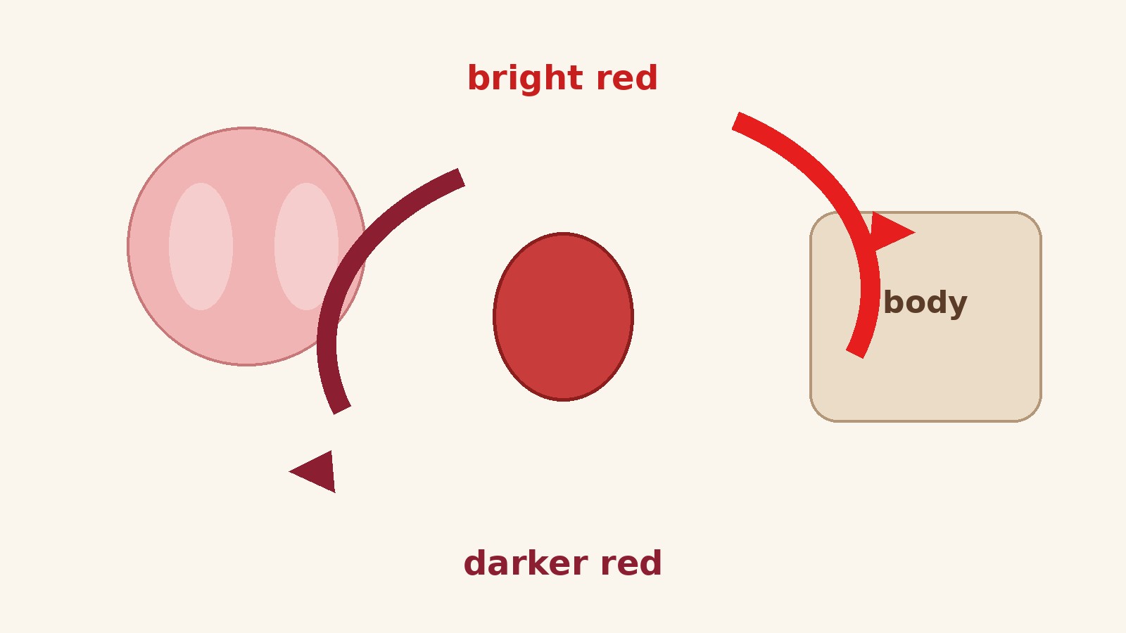
- What changes is the shade. Blood rich in oxygen is bright cherry red. Blood that has delivered its oxygen to the tissues is a darker, duskier red, sometimes described as dark brick red or maroon.
- The blue you see when you look at a vein on the back of your hand is not the color of the blood. It is an optical effect created by the way light travels through skin and is absorbed and scattered before it reaches your eye.
- The red color comes from hemoglobin, the iron containing protein packed into red blood cells. When its iron binds oxygen, the molecule reflects red light strongly. When oxygen leaves, the reflected light shifts and the red darkens.
- This article is general anatomy and physiology information only. It does not diagnose, treat, or advise any individual, and it is not a substitute for professional medical advice.
The Answer in Full
Look at the back of your hand and you will probably see bluish lines running beneath the skin. Cut your finger and the blood that appears is unmistakably red. Both observations are real, and they seem to contradict each other, which is exactly why this question is asked so often. The resolution is simple, even if the physics behind it is not: the blood inside those bluish looking veins is red. It has always been red, and it stays red for the whole journey around the body.
What varies is how red, and how bright. Fresh from the lungs, loaded with oxygen, blood is a vivid, bright red, the color most people picture when they think of blood. After it has traveled through the body and handed its oxygen over to the tissues, it is a much darker red. Cleveland Clinic hematologist Dr. Alan Lichtin puts the range plainly: blood varies from a bright cherry red to a dark brick red, but it is always red. His summary of the whole question is shorter still: “Blood is not blue.”
That single fact clears up the most common version of the myth, the idea that blood is blue while it is inside the body and turns red only when it meets the air. A blood sample drawn from a vein into a sealed tube, with no air contact at all, is dark red, not blue. Blood given by a donor looks dark red in the collection bag for the same reason. The air does not perform a color change. Oxygen does, but oxygen changes red to a brighter red, never blue to red.
Why Blood Is Red: Hemoglobin and Iron
Blood looks red because of what is inside its red blood cells. Red blood cells, also called erythrocytes, are disc shaped cells whose main job is to carry oxygen from the lungs to the tissues and to help carry carbon dioxide back to the lungs for exhalation. The American Red Cross describes red cells as the component that contains hemoglobin and gives blood its red color, and NHS Blood Donation likewise describes red cells as the cells that carry oxygen around the body and carbon dioxide to the lungs to breathe out.
Hemoglobin is the protein that does the carrying. Each hemoglobin molecule is built from protein chains wrapped around pigment groups called heme groups, and at the center of each heme group sits an iron atom. Oxygen binds to that iron. It is this iron and oxygen partnership that produces the color. When oxygen is bound, the hemoglobin molecule absorbs blue green light and reflects red orange light back to the eye, so oxygenated blood looks bright cherry red. When oxygen has been released, the molecule absorbs light differently and reflects a darker, more muted red. Live Science describes the same mechanism in the same terms: hemoglobin bound to oxygen absorbs blue green light and reflects red orange light, and without oxygen connected, blood is a darker red color.
Iron is therefore doing two jobs at once. It is the hook that oxygen hangs on to during transport, and it is the chemical reason the whole fluid looks red. Other oxygen carrying proteins built around different metals produce different colors, which is why some animals famously have blue or green blood, a point covered later in this article. In humans, iron based hemoglobin means red blood, in every shade the molecule can produce.
It is also worth noting what blood is besides red cells. Whole blood is a mixture of red cells, white cells, platelets, and plasma, the pale yellow liquid that carries the cells and dissolved substances around the body. Plasma on its own is not red. Blood looks red because red cells are so numerous and so densely packed with hemoglobin that their color dominates the mixture completely.
Bright Red and Dark Red: The Oxygen Journey
Blood does not sit still inside the body. It circulates in a loop, and its color changes along the way as its oxygen load rises and falls. Following that loop is the clearest way to understand every shade you might encounter.
The loop begins in the lungs. Blood arriving at the lungs is low in oxygen. In the tiny air sacs of the lungs, oxygen passes into the blood and binds to the iron in hemoglobin, forming oxyhemoglobin. This freshly oxygenated blood is bright red. It travels back to the heart, which pumps it out through the arteries to the rest of the body: the brain, the muscles, the skin, the organs. Arterial blood, the blood on the outward leg of the journey, is therefore the bright red kind.
At the tissues, the exchange reverses. Capillaries, the smallest blood vessels, are thin enough for oxygen to pass out of the blood and into the surrounding cells, while carbon dioxide, a waste product of the cells, passes into the blood. As hemoglobin gives up its oxygen, it becomes deoxyhemoglobin, and its color darkens. The blood that collects into the veins for the return leg is therefore darker: a deep, dusky red that people describe as dark brick red, maroon, or burgundy. It is the same fluid, carrying the same hemoglobin, with less oxygen attached.
There is one famous exception to the “arteries carry bright blood, veins carry dark blood” rule, and it is worth knowing because it shows the rule is really about oxygen, not about vessel type. The pulmonary arteries, which carry blood from the heart to the lungs, carry dark, oxygen poor blood, because that blood is on its way to pick oxygen up. The pulmonary veins, which carry blood from the lungs back to the heart, carry bright, oxygen rich blood, because that blood has just collected it. So the color follows the oxygen load, not the name of the vessel. Arteries and veins are defined by the direction of flow relative to the heart, not by the color of their contents.
How much oxygen is unloaded on each pass also varies. Hemoglobin rarely, if ever, gives up all of its oxygen at the tissues, so even venous blood still carries some oxygen. Active muscle takes more oxygen than resting tissue, so blood returning from a hard working muscle is darker than blood returning from a resting area. The scale runs from bright cherry red at full load to deep dark red at low load, and it never leaves the red family.
One more popular number helps convey the pace of this loop. Cleveland Clinic notes that blood completes its round trip, out to the tissues and back, about three times a minute. The exact figure varies with body size and activity, but the picture is right: this is a fast, continuous circulation, and the color shift from bright to dark and back again happens constantly, several times every minute, without you noticing.
Why Veins Look Blue Through Skin
If venous blood is dark red, why do veins look blue? The answer lies in optics, not in blood chemistry. What you see when you look at a vein is not the blood itself. You see light that has traveled into your skin, interacted with tissue and blood, and traveled back out to your eye. That round trip changes which wavelengths survive, and the result looks blue.
White daylight contains all visible wavelengths mixed together. When that light hits skin, different wavelengths behave differently. Red light has a long wavelength, around 700 nanometers, and it penetrates tissue well, reaching 5 to 10 millimeters below the surface, which is where many veins sit. Blue light has a short wavelength, around 475 nanometers, and it is scattered much more easily, so it penetrates only a fraction of a millimeter before bouncing back. This account comes from The Conversation, written with the Australian Red Cross Blood Service, which explains the effect step by step.
Here is what happens over a vein. Red light travels down through the skin, reaches the vein, and is largely absorbed by the hemoglobin in the dark blood there. Relatively little red light makes the return trip to your eye. Blue light, meanwhile, barely penetrates at all: it scatters in the upper layers of skin and bounces back toward your eye in comparatively large amounts. Over ordinary skin, without a vein beneath, you get a mix of reflected wavelengths that reads as skin tone. Over a vein, you get relatively less red and relatively more blue than from the surrounding skin, and your visual system reads that contrast as a blue line. The vein is not blue. Your eye is receiving a blue shifted mixture because the red component was absorbed on the way down and back.
Depth decides how strong the effect is. Very narrow vessels close to the surface, such as the capillary beds that give cheeks and fingertips their pink flush, do not look blue, because the light path is too short for the red absorption to dominate. Deeper veins look bluer, because the longer path removes more red light. The same source notes that vein size matters too, and that the effect varies with skin tone: veins tend to stand out more against very pale skin, which is likely part of the origin of the old phrase “blue blood” for European nobility, people whose untanned skin made their veins conspicuous.
Skin tone, lighting, and vein depth all shift the appearance, which is why veins can look blue on one person, greenish on another, and barely visible on a third, even though the blood inside is the same dark red in all three cases. Greenish veins are the same effect with a slightly different balance of reflected wavelengths, not a different blood color. Nothing about the blood has changed. Only the filter, your skin, and the light have changed.
There is a simple test of this explanation: blood drawn from a blue looking vein fills the tube dark red. The blue was in the viewing conditions, never in the blood.
Why Diagrams Color Veins Blue
If veins are not blue, why does every textbook diagram show them that way? Because diagrams are maps, not photographs. A diagram has to make two different streams easy to tell apart at a glance: the outward stream carrying oxygen to the body and the return stream carrying carbon dioxide back. Red and blue are high contrast, instantly distinguishable colors, and the convention stuck. Generations of students have learned the circulatory system from red and blue diagrams, and the color code, learned early and never corrected, hardens into a belief about the body itself.
The convention is genuinely useful for learning. The problem comes only when the map is mistaken for the territory. A diagram vein is blue for the same reason a road map draws motorways in blue: to distinguish them, not to report their actual color. Once that distinction is clear, the diagram stops being misleading and goes back to being helpful.
The same kind of confusion feeds the phrase “blue blood” for aristocracy. It is a figure of speech, not a medical description, and it probably grew from the visibility of veins through pale, untanned skin, as noted above. Cleveland Clinic makes the same point with a smile: “blue bloods” refers to a noble family lineage or a winning college team, not to the contents of anyone’s veins.
What You Actually See: Cuts, Blood Draws, and Bruises
Everyday encounters with blood confirm the same story, once you know what to look for.
A cut usually bleeds bright red. That is partly because the blood comes from small vessels near the surface, and partly because blood exposed to air picks up oxygen quickly, pushing its hemoglobin toward the bright, fully loaded end of the scale. A deeper cut can bleed darker blood at first, from veins, before it brightens. Either way, the color family is red.
Blood drawn from a vein for a laboratory test looks darker, often surprisingly dark, like red wine or black cherry juice. That is venous blood, low in oxygen, seen without the skin filter. People sometimes worry when they see how dark it is, but dark red is exactly what venous blood is supposed to look like. Blood donation looks the same: the collection bag fills with dark red blood, because donated blood is drawn from a vein.
Bruises tell a longer story. A fresh bruise is reddish or bluish purple, because blood has escaped from damaged vessels into the surrounding tissue, where it sits under skin and is viewed through the same light filtering effect that makes veins look blue. Over days, the trapped blood breaks down, and the bruise passes through green, yellow, and golden brown stages as hemoglobin is converted into other pigments. Those later colors are breakdown products in the tissue, not the color of circulating blood, and they show how hemoglobin chemistry keeps changing after blood leaves the circulation. Dried blood outside the body darkens further still, as hemoglobin breaks down into compounds including methemoglobin and hemichrome, which is why an old bloodstain looks brownish black rather than red.
None of these everyday sights shows blue blood. They show red blood viewed through skin, red blood at different oxygen loads, and red blood breaking down after it has left the vessels. The variety is real, but it is variety within red and its breakdown products, not evidence of a blue stage.
Blood Colors in Other Animals
Human blood is always red, but the animal kingdom runs a wider palette, and the comparison is instructive because it shows exactly what determines blood color: the metal at the heart of the oxygen carrying molecule.
Horseshoe crabs, octopuses, squid, and many other mollusks and arthropods have blue blood. Their oxygen carrier is hemocyanin, a copper containing protein rather than an iron containing one. When hemocyanin binds oxygen, it turns blue, just as hemoglobin turns red when it binds oxygen. Cleveland Clinic notes the same examples: blue blood flows through horseshoe crabs and octopuses because their blood uses the copper containing protein hemocyanin to carry oxygen. Live Science adds that green, clear, and even purple blood appear in other animals, each using a different oxygen carrying molecule.
Green blood appears in some worms and lizards, purple blood in some marine worms, and nearly colorless blood in some Antarctic icefish, which manage with very little hemoglobin at all in cold, oxygen rich water. The fictional Vulcan Spock, who famously bled green in Star Trek, is a nod to the same idea: change the chemistry, and you change the color. None of this affects human blood, but it proves the general rule. Blood color is a property of the oxygen carrier molecule, and in humans that molecule is iron based hemoglobin, so human blood is red.
The comparison also settles a lingering doubt. If blue blood were simply what blood looks like when it is low in oxygen, then low oxygen human blood would be blue, and it is not. The deciding factor is chemistry, not oxygen level alone, and human chemistry gives red.
Rare Human Color Changes
There are rare situations in which human blood takes on an unusual tint, and they are worth mentioning because readers sometimes meet them in news stories and wonder whether they contradict the “always red” rule. They do not contradict it. They are exceptions caused by a changed molecule, not by normal circulation.
One is sulfhemoglobinemia, a rare condition in which a sulfur atom alters the hemoglobin molecule so that blood carries a dark green tint. Cleveland Clinic describes it as typically caused by medications containing sulfur compounds, and notes that the color is temporary: as new red blood cells form, the blood returns to its familiar look. It is a curiosity of hemoglobin chemistry, not a normal shade of blood.
Another is carbon monoxide exposure. Carbon monoxide binds to the iron in heme far more strongly than oxygen does, around 200 times more strongly according to Live Science, and once it is bound it does not let go easily. Blood loaded with carbon monoxide stays bright cherry red, because the molecule is locked in its bright form, and this can make a person look deceptively rosy cheeked even while their blood is failing to deliver oxygen. Again, the color is still in the red family. It is the delivery that has failed, not the color that has changed families.
These cases matter for a different reason too. They show why color alone is a poor guide to what blood is doing. Bright red blood can be carrying carbon monoxide instead of oxygen. Dark red blood can be perfectly normal venous blood. The color tells you something about the hemoglobin molecule, but it does not tell the whole story about oxygen delivery.
A Quick Reference for the Shades
The table below gathers the shades discussed in this article in one place. Every entry is still red, except where a different molecule is involved.
| What you are looking at | Color | Why |
|---|---|---|
| Blood leaving the lungs, in arteries | Bright cherry red | Hemoglobin fully loaded with oxygen, reflecting red orange light |
| Blood returning in veins | Dark brick red, maroon, or burgundy | Hemoglobin partly unloaded, reflecting a darker red |
| A vein seen through skin | Blue or greenish | Light effect: red light absorbed by blood, blue light scattered back by skin |
| A cut at the surface | Bright red | Surface blood, quickly oxygenated on exposure to air |
| Blood drawn from a vein | Dark red, like red wine | Venous blood without the skin filter |
| A fresh bruise | Reddish purple | Blood outside vessels, viewed through skin |
| An older bruise | Green, then yellow, then golden brown | Hemoglobin breaking down into other pigments in tissue |
| Horseshoe crab or octopus blood | Blue | Copper based hemocyanin instead of iron based hemoglobin |
| Rare sulfhemoglobinemia | Dark green | Sulfur altered hemoglobin |
| Blood with carbon monoxide bound | Bright cherry red | Molecule locked in its bright form, oxygen displaced |
Why the Myth Persists
The blue blood myth survives because it is supported from several directions at once. Diagrams color veins blue. Veins genuinely look blue through skin. The phrase “blue blood” is part of everyday language. And the correction, which involves wavelengths, scattering, and absorption, is harder to explain than the myth, which needs only one sentence: blood is blue inside and red outside. Simple, visual, and wrong beats complicated, invisible, and right in most retellings.
Understanding why the myth persists is useful for the same reason understanding the optics is useful: a diagram convention, a trick of light, and a figure of speech can combine into something that feels like personal observation. The veins on your wrist really do look blue. The error is not in your eyes. It is in the inference drawn from what your eyes report.
Frequently Asked Questions
Is blood blue inside your body?
No. Blood inside your body is always red. It ranges from bright cherry red when it is carrying a full load of oxygen to dark brick red or maroon after it has delivered oxygen to the tissues. It is never blue at any point in the circulation.
Does blood turn red when it touches air?
No, not in the sense the myth suggests. Blood does not change from blue to red on contact with air, because it was never blue. Exposure to air can brighten blood, because oxygen in the air binds to hemoglobin and pushes it toward its bright red form, but that is a shift from dark red to bright red, not from blue to red.
Why do my veins look blue if my blood is red?
Because of the way light behaves in skin. Red light penetrates to the vein and is absorbed by the hemoglobin in the blood, while blue light scatters in the upper skin and bounces back to your eye. Over a vein you therefore receive relatively less red and relatively more blue than from the surrounding skin, and the vein reads as blue. The blue is an effect of light and skin, not a property of the blood.
Why do veins sometimes look green?
For the same optical reason, with a slightly different balance of reflected wavelengths, influenced by skin tone, vein depth, and lighting. Greenish veins contain the same dark red blood as blue looking veins. The difference is in the viewing conditions, not in the blood.
Is arterial blood always bright red and venous blood always dark red?
Almost always, but the rule follows oxygen load, not vessel type. The pulmonary arteries carry dark, oxygen poor blood toward the lungs, and the pulmonary veins carry bright, oxygen rich blood away from the lungs. So color tracks how much oxygen the hemoglobin is carrying, and vessel names track the direction of flow relative to the heart.
What color is blood when you donate it?
Dark red. Donated blood is drawn from a vein, so it is blood that has already delivered much of its oxygen to the tissues. Its dark color is normal and expected, and it is the same dark red you would see in any venous blood sample.
Do any animals really have blue blood?
Yes. Horseshoe crabs, octopuses, squid, and many other mollusks and arthropods have blue blood because they use hemocyanin, a copper containing protein, to carry oxygen instead of hemoglobin. Other animals have green, purple, or nearly colorless blood, each color reflecting a different oxygen carrying molecule.
Can human blood ever be green?
Very rarely, in a condition called sulfhemoglobinemia, a sulfur atom alters hemoglobin so that blood carries a dark green tint. It is typically linked to sulfur containing medications and is temporary, resolving as new red blood cells form. It is not a normal shade of circulating blood and it does not mean human blood is sometimes green in the ordinary sense.
Why do diagrams show veins in blue?
As a labeling convention. Red and blue are easy to distinguish, so diagrams use red for the oxygenated outward stream and blue for the deoxygenated return stream. The colors mark the two halves of the loop for teaching purposes. They are not claims about the actual color of the blood.
Does dark blood in a blood test mean something is wrong?
Not on its own. Blood drawn from a vein is supposed to be dark red, because it is returning blood that has given up much of its oxygen. Color alone does not diagnose anything, and laboratory interpretation rests on measured values and the broader clinical picture, not on the shade in the tube.
Sources & references
- Cleveland Clinic, "What Color Is Blood Inside of Your Body?" https://health.clevelandclinic.org/what-color-is-blood (opened and read 26 Oct 2026; hematologist Dr. Alan Lichtin on blood always ranging from bright cherry red to dark brick red, the optical illusion behind blue looking veins, blue blood in horseshoe crabs and octopuses via hemocyanin, and sulfhemoglobinemia).
- Live Science, "Blood in Your Veins Is Not Blue: Here's Why It's Always Red" https://www.livescience.com/62724-blood-in-your-veins-not-blue.html (opened and read 26 Oct 2026; hemoglobin and heme absorbing blue green light and reflecting red orange light, carbon monoxide binding around 200 times more strongly than oxygen, blue light penetrating tissue less far than red light, green and purple blood in other animals, dried blood darkening via methemoglobin and hemichrome).
- NHS Blood Donation, "What's in your blood" http://www.blood.co.uk/why-give-blood/whats-in-your-blood/ (opened and read 26 Oct 2026; red cells carry oxygen around the body and carbon dioxide to the lungs, plasma carries the cells, platelets help blood clot).
- The Conversation via ScienceAlert, "Why Do Our Veins Often Look Blue if Blood Is Actually Red?" https://www.sciencealert.com/why-are-white-people-s-veins-blue-when-blood-is-red (opened and read 26 Oct 2026; red light reaching 5 to 10 millimeters below the skin, blue light around 475 nanometers scattering within a fraction of a millimeter, vein depth and skin tone effects, "blue blood" phrase origin).


