Short-answer box
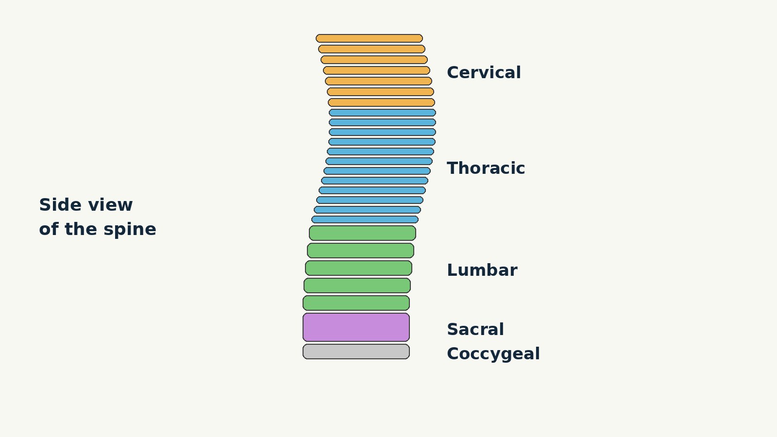
The spine, also called the vertebral column or spinal column, is a flexible column of bones, discs, ligaments, and muscles that supports the head and body, allows movement, and protects the spinal cord. It has five regions, from the neck to the base of the spine: cervical, thoracic, lumbar, sacral, and coccygeal. The column develops as 33 vertebrae. In an adult, 24 vertebrae stay separate and movable, while the sacral vertebrae fuse into one bone called the sacrum and the coccygeal vertebrae fuse into one bone called the coccyx, so the adult column has 26 bones in total.
Each movable vertebra has a solid body at the front that carries weight, an arch at the back that forms a protective ring, and projections where muscles and ligaments attach. Between most movable vertebrae sits an intervertebral disc that cushions the bones and allows small movements. The spinal cord runs through a canal formed by the stacked vertebrae, and 31 pairs of spinal nerves leave the column to carry signals between the brain and the rest of the body. From the side, the adult spine has four gentle curves that help it stay strong, flexible, and able to absorb shock.
What the spine is
Think of the spine as a stack of building blocks that can bend, twist, and spring back. The blocks are the vertebrae. The pads between many of the blocks are the intervertebral discs. The joints between the blocks, the ligaments that tie the blocks together, and the muscles that pull on the blocks all work together to make a structure strong enough to hold the body upright and flexible enough to let a person bend forward, lean back, turn, and reach.
The spine has three main jobs. First, it supports the weight of the head and upper body and passes that weight down toward the pelvis and legs. Second, it protects the spinal cord, the main bundle of nerve tissue connecting the brain to the rest of the body. Third, it allows movement. No single joint in the spine moves very far on its own, but the small movements at many joints add up to a large range of motion for the head, neck, and trunk.
The five regions of the spine
The vertebral column is divided into five regions, named from the top down: cervical, thoracic, lumbar, sacral, and coccygeal. Vertebrae in each region are named for that region and numbered from the top down, which gives every level a short address. A diagram can point to one level, such as C4 or L5, and a reader who knows the regions will know at once where that level sits.
| Region | Where it is | Vertebrae | Adult bones |
|---|---|---|---|
| Cervical | Neck | 7, named C1 to C7 | 7 separate vertebrae |
| Thoracic | Upper and middle back | 12, named T1 to T12 | 12 separate vertebrae |
| Lumbar | Lower back | 5, named L1 to L5 | 5 separate vertebrae |
| Sacral | Between the hip bones | 5 fused sacral vertebrae | 1 bone, the sacrum |
| Coccygeal | Base of the spine | Usually 4 fused coccygeal vertebrae, sometimes 3 or 5 | 1 bone, the coccyx |
Adding the developmental counts gives 7 plus 12 plus 5 plus 5 plus 4, which equals 33 vertebrae. Adding the adult bones gives 7 plus 12 plus 5 plus 1 plus 1, which equals 26 bones. The difference is fusion, not missing bones. The sacral and coccygeal vertebrae join together during growth, and fusion is usually complete in adult life. Vertebrae change shape by region to match their job.
Cervical region: the neck, C1 to C7
The cervical region has seven vertebrae, named C1 to C7, and forms the neck. The top vertebra, C1, is called the atlas, after the figure in Greek myth pictured supporting the heavens, because it supports the skull. Unlike a typical vertebra, the atlas has no body and no spinous process. It is ring shaped, with a front arch and a back arch, and its upper joint surfaces meet the base of the skull. The second vertebra, C2, is called the axis, because it serves as the axis for rotation when the head turns. It has a bony projection called the dens that extends upward and joins the front arch of the atlas, held in place by a ligament.
Typical cervical vertebrae, such as C4 or C5, are small, with a small body for a light load. Their spinous processes are usually split into two points, a shape described as bifid, except for C7, whose spinous process is much longer and can be felt as a bump at the base of the neck. Each cervical transverse process has an opening called a transverse foramen, and the vertebral arteries, which supply the brain, pass upward through these openings.
Thoracic region: the upper and middle back, T1 to T12
The thoracic region has 12 vertebrae, named T1 to T12, and forms the upper and middle back. Thoracic vertebrae are larger than cervical vertebrae, and their bodies grow larger as the column descends, because the load they carry grows. A typical middle thoracic vertebra has a long spinous process that angles downward and overlaps the vertebra below it.
Thoracic vertebrae are also the only vertebrae that join directly to ribs. Each one has joint surfaces on the sides of its body, called costal facets, for the head of a rib, and an additional facet on the transverse process for the tubercle of a rib. The word costal means relating to the ribs. The rib cage, thoracic vertebrae, and breastbone form a protective cage around the chest organs, and the rib joints allow the chest to expand and narrow during breathing.
Lumbar region: the lower back, L1 to L5
The lumbar region has five vertebrae, named L1 to L5, and forms the lower back. Lumbar vertebrae carry the greatest amount of body weight, so they have a large, thick vertebral body, short transverse processes, and a short, blunt spinous process that projects straight back. Their large joint surfaces, with the upper process facing backward and the lower process facing forward, guide and limit movement in the lower back.
The lumbar region allows a large share of bending forward and leaning back, and it transfers the weight of the upper body to the sacrum and pelvis below. Its discs are the thickest in the column, so the lumbar region can also absorb a large share of the shock from walking, lifting, and other everyday movements. The parts that carry the most weight are built the most heavily, in both body size and disc thickness.
Sacral region: the sacrum, formed from five fused vertebrae
The sacrum is a triangular bone at the base of the spine, formed by the fusion of five sacral vertebrae. It is thick and wide across its upper base, where it bears weight, and it tapers to a lower tip that does not bear weight in standing. On the front surface of an older adult sacrum, the lines where the vertebrae fused can still be seen as four ridges. On the back surface, a bumpy middle ridge called the median sacral crest is the remnant of the fused spinous processes, and side ridges called lateral sacral crests are the remnant of the fused transverse processes.
The sacrum sits between the two hip bones and joins the ilium portion of each hip bone to form the sacroiliac joints, which are immobile joints of the pelvis. That connection lets the spine pass weight into the pelvis and then into the legs. A bony tunnel called the sacral canal passes down through the sacrum, and paired openings called sacral foramina allow branches of the sacral spinal nerves to leave the sacrum. The upper joint surfaces of the sacrum meet the lower joint surfaces of L5, the last movable vertebra above it.
Coccygeal region: the coccyx, or tailbone
The coccyx, commonly called the tailbone, is formed by the fusion of four very small coccygeal vertebrae, though the number is sometimes three or five. It sits at the very base of the spine, below the sacrum, and joins the lower tip of the sacrum at a slightly movable joint. The coccyx is not weight bearing when a person is standing, but it can receive some body weight when a person is sitting, and it serves as an attachment point for muscles and ligaments of the pelvic floor. As an anchor at the base of the pelvis, it helps tie together the soft tissues that support the pelvic organs and close the bottom of the pelvis.
Inside a typical vertebra
Most vertebrae follow the same basic pattern: a body, a vertebral arch, and seven processes. The first two cervical vertebrae are exceptions, as described above, and the sacrum and coccyx are fused versions of the pattern, but the pattern still explains what to look for in any region.
The vertebral body is the front portion of a vertebra and the part that supports body weight. It is a short, roughly cylindrical block of bone, and the bodies of neighboring vertebrae are separated and strongly united by an intervertebral disc. Because the load grows as the column descends, vertebral bodies increase in size and thickness from the neck to the lower back. The body is the strongest part of a vertebra for carrying a vertical load, but it is not the part that protects the cord. That job belongs to the arch behind it.
The vertebral arch forms the back portion of each vertebra. It is made of four parts: a right and a left pedicle, and a right and a left lamina. Each pedicle is anchored to the back side of the vertebral body and forms one side wall of the arch. Each lamina forms part of the back roof of the arch. Together, the body and the arch enclose a large opening called the vertebral foramen. When the vertebrae are stacked, these openings line up to form a continuous tunnel called the vertebral canal, also called the spinal canal, through which the spinal cord passes. Where two neighboring vertebrae meet, notches in their pedicles line up to form a smaller side opening called an intervertebral foramen, which is the exit route for a spinal nerve.
Seven processes project from the vertebral arch. Two transverse processes project sideways, one on each side, from the junction of the pedicle and the lamina. The single spinous process projects backward at the midline of the back, and the spinous processes are the bumps felt as a row down the middle of the back. Transverse and spinous processes serve mainly as attachment sites for muscles and ligaments. Four articular processes form the joints between neighboring vertebrae: on each side, a superior articular process faces upward and an inferior articular process faces downward, and their contact surfaces, called facets, form slightly movable facet joints. The shape and orientation of those surfaces vary by region and play a major role in deciding what kind of movement each region allows.
Intervertebral discs: the pads between vertebrae
Between the bodies of adjacent vertebrae sits an intervertebral disc, a pad of fibrocartilage that fills the gap between the bones, unites them strongly, and provides padding during weight bearing. It is anchored to the bodies above and below it, and it changes shape as the vertebrae move, which lets the column bend while staying connected.
Each disc has two main parts. The outer layer, called the annulus fibrosus, is a tough, fibrous ring anchored to the outer margins of the neighboring vertebral bodies. The inner part, called the nucleus pulposus, is a softer, gel-like material with a high water content that resists compression and is important for weight bearing. A thin layer of cartilage, called an endplate, sits between the disc and each vertebral body and helps anchor the disc to the bone.
The adult spine has 23 intervertebral discs: 6 cervical, 12 thoracic, and 5 lumbar. There is no disc between C1 and C2, and the lowest disc sits between L5 and the sacrum. There are no discs between the fused vertebrae of the sacrum or coccyx. Together, the discs make up about one quarter of the length of the column from the base of the skull to the top of the pelvis. Discs are thin in the cervical region and thickest in the lumbar region. When a person bends forward, the front of a disc is compressed and the back expands, and the pattern reverses when leaning back. The movement at any one disc is small, but the small movements at 23 discs add up to the large movements of the whole trunk. With increasing age, the water content of the nucleus pulposus gradually declines and the disc becomes thinner and less flexible.
The spinal cord and nerves, in plain language
The spinal cord is a long, roughly cylindrical bundle of nerve tissue that runs through the vertebral canal. It is part of the central nervous system, together with the brain, and it serves as the main pathway for signals traveling between the brain and the rest of the body.
The cord begins at the base of the brain and runs down through the canal formed by the vertebrae. In an adult, the cord itself ends near the level of the first or second lumbar vertebra, at a tapered tip called the conus medullaris. The cord is therefore shorter than the vertebral column. Below that tip, bundles of nerve roots continue down the canal and look, as a group, like a horse tail. That bundle is called the cauda equina, from the Latin words for horse tail.
Inside the cord, gray matter made mostly of nerve cell bodies forms a shape like the letter H or a butterfly in the middle of the cord, while white matter made mostly of nerve fibers surrounds it and carries signals up and down the cord. The gray matter has rear horns called dorsal horns, front horns called ventral horns, and, in the thoracic and lumbar regions, side horns called lateral horns. A small central canal runs through the gray matter and is filled with cerebrospinal fluid, the fluid that surrounds the brain and spinal cord. The cord is covered by three membranes called the meninges: tough dura mater outside, web-like arachnoid mater in the middle, and delicate pia mater inside. Cerebrospinal fluid between the arachnoid and pia layers cushions the cord.
From the spinal cord branch 31 pairs of spinal nerves, one pair at most levels, with one nerve leaving on each side of the column: 8 cervical, 12 thoracic, 5 lumbar, 5 sacral, and 1 coccygeal. Each spinal nerve leaves through an intervertebral foramen, the side opening formed by two neighboring vertebrae, except the sacral nerves, which leave through the sacral foramina. The first cervical pair is an exception: it leaves between the base of the skull and the atlas, because there are eight cervical nerve pairs but only seven cervical vertebrae. Each spinal nerve is formed by two roots that join just outside the cord. The rear root, called the dorsal root, carries sensory information toward the central nervous system, such as touch, temperature, and position. The front root, called the ventral root, carries motor information away from the central nervous system toward muscles and glands. The swelling on the dorsal root, called the dorsal root ganglion, holds the cell bodies of the sensory nerve cells. Once the roots join, the nerve is a mixed nerve, carrying both sensory and motor fibers.
After leaving the column, spinal nerves branch into smaller nerves for specific areas of skin and muscle: cervical nerves serve the neck, shoulders, and arms, thoracic nerves serve the trunk, and lumbar and sacral nerves serve the pelvis and legs.
Curves of the spine
Seen from the front or back, a healthy adult spine looks fairly straight. Seen from the side, it has four gentle curves along its length, and those curves are part of what makes the spine strong, flexible, and able to absorb shock. When the load on the spine increases, for example when a person carries a heavy backpack, the curves deepen slightly to take up the extra weight, and they spring back when the weight is removed.
The four curves fall into two kinds. Primary curves are kept from the original curve of the fetus, when the body is curled forward. In the adult, that early curve remains as the thoracic curve and the sacrococcygeal curve, formed by the sacrum and coccyx. Those curves are concave toward the front of the body. Secondary curves develop after birth, as a child learns to sit upright, stand, and walk, and they curve in the opposite direction, concave toward the back. The cervical curve develops as an infant begins to hold the head upright while sitting. The lumbar curve develops later, as the child begins to stand and walk. In adults, the lumbar curve is generally deeper in females than in males, a normal variation.
From the side, the curves give the adult spine a gentle S shape: the neck curves slightly inward at the front, the upper back curves slightly outward at the back, the lower back curves inward at the front again, and the sacrum and coccyx curve outward at the back. Three terms describe curves that are larger than usual or in an unusual direction: kyphosis is an excessive backward curve of the thoracic region, lordosis is an excessive forward curve of the lumbar region, and scoliosis is a sideways curve, usually with some twisting. Those words describe shape, not a single cause.
Ligaments: the ties that hold the column together
Ligaments are tough bands of connective tissue that join bone to bone. In the spine, ligaments run along the length of the column, tie neighboring vertebrae together, and resist movements that would go too far. They do not produce movement on their own. They limit it, guide it, and help the column spring back after bending.
The anterior longitudinal ligament runs down the front side of the entire vertebral column, uniting the front sides of the vertebral bodies and the discs between them, and it resists excessive backward bending. The posterior longitudinal ligament runs inside the vertebral canal, on the back sides of the vertebral bodies and in front of the spinal cord, and it helps support the column when bending forward. The ligamentum flavum, whose name means yellow ligament, sits behind the spinal cord inside the canal. It is a series of short, paired ligaments, each joining the laminae of two neighboring vertebrae, and its many elastic fibers give it a yellow color and let it stretch when the spine bends and pull back when the spine straightens.
On the back of the column, the supraspinous ligament joins the spinous processes of the thoracic and lumbar vertebrae and supports the column during forward bending. In the neck, where the cervical spinous processes are short, that ligament expands into a stronger sheet called the nuchal ligament, which attaches to the cervical spinous processes and extends upward and backward to the base of the skull, where it helps support the skull and keeps the head from falling forward. Smaller ligaments join neighboring spinous and transverse processes. The facet joints are held together by their own capsular ligaments.
Muscles: the motors and stabilizers
Muscles produce the movements that ligaments only limit. Muscles attach to the transverse and spinous processes of vertebrae, to the ribs, to the pelvis, and to the skull, and when a muscle shortens, it pulls on those attachment points and moves the part it is attached to. Muscles also hold posture: even when a person is standing still, back and abdominal muscles are working at a low level to keep the trunk upright and the curves of the spine in their resting shape.
The deep muscles of the back lie closest to the vertebral column, on each side of it, and span the full length from the skull to the pelvis. They include the erector spinae group, a large set of muscles that run lengthwise along the back and extend the spine, and the deeper transversospinal muscles, which run from the transverse process of one vertebra to the spinous process of a vertebra above it and help with extension, side bending, and rotation. The deepest and shortest muscles span only one or two joints and make small adjustments to the position of individual vertebrae. Together, those layers give the spine both power for large movements and fine control for small ones.
Muscles in front of the spine matter just as much as muscles behind it. The abdominal muscles at the front of the trunk work with the back muscles to bend, brace, and steady the trunk. The balance between muscle groups keeps the column stable: back muscles pull the trunk backward, abdominal muscles pull it forward, and side muscles pull it to each side.
A note on health questions
If you have a health concern about your spine, speak with a qualified health professional.
Frequently asked questions
How many bones are in the spine?
The adult spine has 26 bones: 24 separate vertebrae plus the sacrum and the coccyx. It develops as 33 vertebrae, with fusion accounting for the difference.
What are the five regions of the spine?
From the top down: cervical (7 vertebrae), thoracic (12), lumbar (5), sacral (1 fused bone from 5 vertebrae), and coccygeal (1 fused bone from usually 4 vertebrae).
What is the difference between the sacrum and the coccyx?
The sacrum is a large triangular bone that joins the spine to the pelvis and passes weight into the hip bones. The coccyx is a much smaller anchor bone at the base of the spine for pelvic muscles and ligaments.
What are the main parts of a single vertebra?
A typical vertebra has a weight bearing body at the front, an arch at the back made of two pedicles and two laminae, and seven processes: two transverse, one spinous, and four articular. Body and arch together enclose the vertebral foramen, through which the cord passes.
How many intervertebral discs are there?
There are 23 discs: 6 cervical, 12 thoracic, and 5 lumbar. There is no disc between C1 and C2, or between fused sacral or coccygeal vertebrae. Each disc has a tough outer ring, the annulus fibrosus, and a gel-like center, the nucleus pulposus.
Does the spinal cord run the whole length of the spine?
No. The cord ends near the first or second lumbar vertebra, at a tip called the conus medullaris. Below it, nerve roots called the cauda equina continue down the canal. The column is longer than the cord.
How many pairs of spinal nerves are there?
There are 31 pairs: 8 cervical, 12 thoracic, 5 lumbar, 5 sacral, and 1 coccygeal. Each pair leaves through an intervertebral foramen, except the first cervical pair, which leaves between the skull base and the atlas, and the sacral pairs, which leave through the sacral foramina. Each nerve carries sensory and motor signals.
Why does the spine have curves?
The four curves, cervical, thoracic, lumbar, and sacrococcygeal, increase strength and flexibility and help absorb shock. The thoracic and sacrococcygeal curves are kept from before birth, while the cervical and lumbar curves develop as a child learns to sit, stand, and walk. Together they give the side view a gentle S shape.
What do spinal ligaments do?
Ligaments join bone to bone and limit bending and twisting. The anterior longitudinal ligament runs along the front, the posterior longitudinal ligament runs inside the canal, the ligamentum flavum joins neighboring laminae, and the supraspinous ligament joins the spinous processes.
What muscles support the spine?
Deep back muscles, including the erector spinae group and deeper transversospinal muscles, extend the spine, help with side bending and rotation, and stabilize joints. Abdominal muscles work with them to bend, brace, and steady the trunk.
Sources & references
- OpenStax, Anatomy and Physiology 2e, Chapter 7.3, The Vertebral Column. https://open.oregonstate.education/aandp/chapter/7-4-the-vertebral-column/
- U.S. National Library of Medicine, MedlinePlus Medical Encyclopedia, Spinal fusion series, Normal anatomy. https://medlineplus.gov/ency/presentations/100121_1.htm
- U.S. National Library of Medicine, National Center for Biotechnology Information Bookshelf, StatPearls, Anatomy, Back, Intervertebral Discs. https://www.ncbi.nlm.nih.gov/books/NBK470583/
- U.S. National Library of Medicine, National Center for Biotechnology Information Bookshelf, StatPearls, Anatomy, Back, Muscles. https://www.ncbi.nlm.nih.gov/books/NBK537074/?report=reader
- Chabot College, LibreTexts, Spinal Cord Anatomy, Section 8.6. https://med.libretexts.org/Courses/Chabot_College/BIOS15%3A_Lab_Manual/08%3A_Nervous_System/8.06%3A_Spinal_Cord_Anatomy


