Short answer
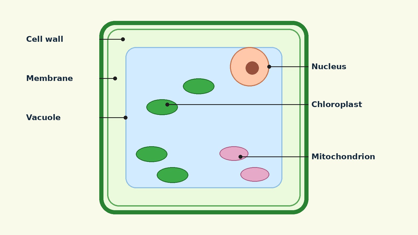
- A labeled plant cell diagram shows a plant cell from the outside inward. The outermost layer is the cell wall, with the cell membrane just inside it. Inside the cell are the nucleus, chloroplasts, mitochondria, ribosomes, endoplasmic reticulum, Golgi apparatus, cytoplasm, and a large central vacuole. The three features that most clearly mark the diagram as a plant cell, rather than an animal cell, are the cell wall, the chloroplasts, and the large central vacuole. Each label is more than a name to memorize. The shape and position of each part reflect the job it does.
How to Read the Diagram
Start at the outside and work inward. The cell wall is the outermost line. The cell membrane, also called the plasma membrane, is the thinner line just inside the wall. Next find the largest interior space: the large central vacuole, usually drawn as a clear, pale region taking up much of the cell. Then look for the nucleus, often a large round body with a darker spot inside it, the nucleolus. Chloroplasts are usually green ovals in the cytoplasm around the vacuole. Mitochondria are smaller ovals with folded inner lines. Ribosomes are tiny dots, the endoplasmic reticulum is a folded membrane network, and the Golgi apparatus is a stack of curved sacs. Plasmodesmata, when shown, are tiny channels crossing the cell wall.
A diagram is a simplification. Real plant cells vary by tissue, age, and function. A leaf cell that carries out photosynthesis looks different from a root cell that absorbs water, and a young cell with several small vacuoles looks different from a mature cell with one large central vacuole. Most labeled diagrams show a generalized photosynthetic plant cell. Treat it as a map of the common parts, not as a photograph of every plant cell. Also, do not judge importance by size alone: ribosomes are tiny in a diagram, but protein synthesis depends on them.
Labeled Diagram at a Glance
| Label | Where to find it | Main job |
|---|---|---|
| Cell wall | Outermost layer | Protects the cell and gives shape and support |
| Cell membrane | Just inside the cell wall | Controls what passes into and out of the cell |
| Cytoplasm | Between membrane and nuclear envelope | Holds organelles and hosts many reactions |
| Nucleus | Large round body, often near the edge | Houses DNA and directs ribosome and protein synthesis |
| Chloroplast | Green ovals in cytoplasm | Carries out photosynthesis |
| Large central vacuole | Largest interior compartment | Stores water and helps maintain turgor |
| Mitochondrion | Small oval with folded inner membrane | Produces most cell ATP through respiration |
| Ribosome | Tiny dots, free or on rough ER | Builds proteins from messenger RNA |
| Rough ER | Membrane network studded with ribosomes | Makes and modifies proteins |
| Smooth ER | Membrane network without ribosomes | Makes lipids, with roles that vary by cell type |
| Golgi apparatus | Stack of flattened sacs | Modifies, sorts, and packages proteins and lipids |
| Plasmodesmata | Channels through the cell wall | Connect neighboring cells for transport |
Cell Wall
The cell wall is the rigid outer covering of a plant cell, drawn as the outermost boundary. Its major organic molecule in plants is cellulose, a polysaccharide made of glucose units. The wall protects the cell, provides structural support, and gives the cell its shape. Plant cells often look boxy or rectangular in diagrams for this reason, while animal cells, which lack a wall, are drawn with more flexible outlines.
The wall also limits how much the cell can swell when water enters. Water entry therefore builds turgor pressure, covered in the vacuole section, rather than bursting the cell. A common error is to treat the cell wall and the cell membrane as the same line. In a careful diagram they are two lines: the wall outside, the membrane inside.
Cell Membrane
The cell membrane is also called the plasma membrane. In a diagram it is the line just inside the cell wall, drawn much thinner than the wall. Its thinness should not be mistaken for unimportance. The membrane is the living boundary of the cell.
Structurally, the plasma membrane is a phospholipid bilayer with embedded proteins: a double layer of lipid molecules with proteins set into it. Functionally, it controls the passage of organic molecules, ions, water, and oxygen into and out of the cell. Wastes such as carbon dioxide and ammonia also leave through it. The membrane is selective rather than simply open or closed, and transport proteins help move substances that cannot cross the lipid layer on their own. The wall gives rigid support. The membrane controls exchange.
Cytoplasm
The cytoplasm is the entire region between the plasma membrane and the nuclear envelope. It consists of organelles suspended in a gel-like material called cytosol, plus the cytoskeleton and various chemicals. The pale background inside the cell is not empty space. It is cytoplasm, and many metabolic reactions take place there, including protein synthesis.
The cytosol is mostly water. OpenStax Biology 2e describes cytoplasm as 70 to 80 percent water, with a semi-solid consistency that comes from the proteins within it. Simple sugars, polysaccharides, amino acids, nucleic acids, and fatty acids are also present, along with ions such as sodium, potassium, and calcium. The cytoskeleton, a network of protein fibers, helps maintain cell shape, secures some organelles in position, and allows cytoplasm and vesicles to move. If a label points to an apparently blank area between organelles, it is usually pointing to cytoplasm or cytosol.
Nucleus
The nucleus is typically the most prominent organelle in a cell, usually drawn as a large round or oval body. It houses the cell DNA and directs the synthesis of ribosomes and proteins. A more precise description than control center is this: the nucleus stores genetic instructions and controls access to them, while much of the actual work of making proteins happens at ribosomes in the cytoplasm.
The nucleus is bounded by the nuclear envelope, a double membrane made of an inner and an outer phospholipid bilayer. The envelope has nuclear pores, which control the passage of ions, molecules, and RNA between the nucleoplasm inside the nucleus and the cytoplasm outside it. Chromatin, material made of DNA plus proteins, forms chromosomes, which are only clearly visible when the cell is getting ready to divide.
Inside the nucleus, look for a darker, denser spot: the nucleolus, a condensed chromatin region where ribosome synthesis occurs. The nucleolus aggregates ribosomal RNA with associated proteins to assemble ribosomal subunits, which are then transported out through the pores to the cytoplasm. A mature plant cell diagram may show the nucleus pushed to one side by the large central vacuole. That is an effect of the vacuole, not an error.
Chloroplast
Chloroplasts are the green, disc-like organelles in a plant cell diagram, and they are the reason the cell can carry out photosynthesis. Photosynthesis is the series of reactions that use carbon dioxide, water, and light energy to make glucose and oxygen. Light energy captured in chloroplasts becomes chemical energy stored in sugars, and those sugars can then be used in cellular respiration in mitochondria to produce ATP.
A chloroplast has outer and inner membranes. Within the inner membrane is a set of stacked, fluid-filled membrane sacs called thylakoids. Each thylakoid stack is a granum, plural grana. The fluid surrounding the grana is the stroma. Light harvesting reactions take place in the thylakoid membranes, and sugar synthesis takes place in the stroma. Chloroplasts also contain chlorophyll, the green pigment that captures light energy, and they have their own DNA and ribosomes.
A simple diagram may draw each chloroplast as a plain green oval. A detailed diagram may show thylakoids, grana, and stroma inside the oval. Both are correct at different levels of detail. The green oval as a whole is the chloroplast, while thylakoids and grana are structures inside it. Not every plant cell has chloroplasts: root cells may lack them or contain other plastids instead.
Large Central Vacuole
The large central vacuole is the largest compartment in a mature plant cell diagram. It is a membrane-bound sac that occupies most of the volume of the cell, and its membrane is called the tonoplast. In a diagram it is usually drawn as a large, clear region in the middle of the cell, with other organelles arranged around it in a thin layer of cytoplasm.
The vacuole acts as a storage compartment and a water reservoir, and it is a site where macromolecules are broken down. It also regulates the concentration of water in the cell under changing conditions. When the central vacuole holds more water, the cell becomes larger without having to invest considerable energy in synthesizing new cytoplasm. That expansion supports growth and helps keep the cell wall supported from inside.
That support is called turgor. When water leaves the central vacuole and cytoplasm, the vacuole shrinks and leaves the cell wall unsupported, which produces the wilted appearance of a plant that has not been watered. Young plant cells often have several smaller vacuoles that later merge into a large central vacuole, so a young cell diagram may show multiple vacuoles while a mature cell diagram shows one.
Mitochondria
Mitochondria are the oval organelles with folded inner membranes in a plant cell diagram. The singular is mitochondrion. They are often called the powerhouses of the cell because they make adenosine triphosphate, or ATP, the main energy-carrying molecule of the cell. Cellular respiration is the process of making ATP using the chemical energy in glucose and other nutrients. In mitochondria, this process uses oxygen and produces carbon dioxide as a waste product.
Each mitochondrion is a double membrane organelle. The inner membrane has folds called cristae, which increase its surface area. The space between the two membranes is the intermembrane space, and the space inside the inner membrane is the mitochondrial matrix. ATP synthesis takes place on the inner membrane. Mitochondria also have their own ribosomes and DNA, a feature they share with chloroplasts.
Chloroplasts and mitochondria are partners in energy flow, not rivals. Chloroplasts capture light energy and store it in sugars. Mitochondria release energy from sugars in a form the cell can use, ATP, which powers processes throughout the cell. In a diagram, mitochondria are usually smaller than chloroplasts and not green. Their folded inner lines are the visual clue.
Ribosomes
Ribosomes are the smallest labeled structures in most plant cell diagrams, often drawn as tiny dots. Their small size hides a central job: ribosomes are the cellular structures responsible for protein synthesis. They receive instructions in the form of messenger RNA, or mRNA, transcribed from DNA in the nucleus. The mRNA travels to ribosomes, which translate the code in the mRNA into a specific order of amino acids in a protein. Amino acids are the building blocks of proteins.
A ribosome consists of two subunits, large and small, and is a large complex of protein and RNA. Ribosomes may float freely in the cytoplasm or be attached to the cytoplasmic side of the endoplasmic reticulum or the outer membrane of the nuclear envelope. Through an electron microscope they appear as clusters called polyribosomes or as single, tiny dots. In a textbook diagram they are simplified to dots.
The location of a ribosome hints at the destination of the protein it makes. Free ribosomes generally make proteins that will function in the cytoplasm. Ribosomes attached to the rough endoplasmic reticulum make proteins that enter the endomembrane system, for secretion, for membranes, or for other compartments. A diagram that shows only a few dots is using dots as a symbol for a much larger population.
Endoplasmic Reticulum
The endoplasmic reticulum, often shortened to ER, is drawn as a folded network of membranes near the nucleus. It is part of the endomembrane system, a group of membranes and organelles that works together to modify, package, and transport lipids and proteins. The system includes the nuclear envelope, the endoplasmic reticulum, the Golgi apparatus, lysosomes, and vesicles, and it interacts closely with the plasma membrane.
There are two types of ER, and a careful diagram labels them separately. Rough endoplasmic reticulum, or RER, is studded with ribosomes on its cytoplasmic surface, which gives it a dotted outline in diagrams. It is the site of protein synthesis and modification for proteins entering the endomembrane system, and membrane and secretory proteins are synthesized there. Smooth endoplasmic reticulum, or SER, lacks ribosomes and therefore looks smooth. It is involved in lipid synthesis, carbohydrate metabolism, detoxification of medications and poisons in relevant cell types, and storage of calcium ions.
The nuclear envelope is continuous with the ER, which is why the ER is usually drawn connected to the nucleus. Proteins made on rough ER can be modified in the ER, then carried in vesicles to the Golgi apparatus. Follow that flow when reading a diagram: nucleus to rough ER to Golgi apparatus is one of the main production lines of the cell. Use the dots as your guide: dotted membrane is rough ER, smooth membrane without dots is smooth ER.
Golgi Apparatus
The Golgi apparatus, also called the Golgi body or Golgi complex, is drawn as a stack of flattened, curved sacs, often near the endoplasmic reticulum. The sacs are called cisternae. The stack usually has a receiving side facing the ER and a shipping side facing away from it. Proteins and lipids arriving from the ER enter the receiving side, are modified as they pass through the stack, and leave the shipping side in vesicles bound for their destinations.
The Golgi apparatus modifies, sorts, tags, packages, and distributes lipids and proteins. Modification can include adding carbohydrate groups to proteins. Sorting means directing different cargoes to different destinations: some to the plasma membrane, some for secretion outside the cell, and some to other compartments within the cell. Packaging means enclosing the finished cargo in a membrane vesicle so it can travel without mixing with the rest of the cytoplasm.
In plant cells, the Golgi apparatus also contributes materials for the cell wall and for the plasma membrane during growth and division. Do not confuse it with the endoplasmic reticulum. The ER is a continuous folded network, often drawn as a maze. The Golgi apparatus is a discrete stack of separate sacs. That visual difference, a network versus a stack, is the most reliable way to tell the two labels apart.
Plasmodesmata
Plasmodesmata are the tiny channels that pass through the cell walls of adjacent plant cells. The singular is plasmodesma. In a detailed diagram they appear as small gaps or bridges in the cell wall, sometimes drawn as a thin line connecting the cytoplasm of one cell to the cytoplasm of its neighbor. In a simple single cell diagram they may be omitted entirely, or shown only as short marks on the wall. Their absence from a simple diagram does not mean they are unimportant.
Plasmodesmata connect the cytoplasm of neighboring cells and enable materials to be transported from cell to cell, and thus throughout the plant. That connection matters because the cell wall separates neighboring plant cells. Without channels through the wall, each cell would be isolated behind its own rigid covering. With plasmodesmata, water, nutrients, and signaling materials can move directly between cells, allowing tissues to coordinate growth.
When labeling plasmodesmata, be precise about location. They are not organelles floating in the cytoplasm, and they are not part of the cell membrane alone. They are structures of the wall region, channels that cross it. If a diagram shows only one plant cell, the label will point to the wall edge, where a neighboring cell would sit in a tissue drawing.
Peroxisomes and Other Plastids
Some detailed diagrams include small round sacs labeled peroxisomes. Peroxisomes are small, round organelles enclosed by single membranes. They carry out oxidation reactions that break down fatty acids and amino acids, and they detoxify many poisons that may enter the cell. Many of these reactions release hydrogen peroxide, which would be damaging to cells, but when the reactions are confined to peroxisomes, enzymes safely break down the hydrogen peroxide into oxygen and water. Plant cells contain specialized peroxisomes called glyoxysomes, which convert stored fats into sugars.
Other diagrams may label plastids more generally. Chloroplasts are one type of plastid. Other plastids store starch or pigments, and they help explain why some plant tissues are colorful or starchy even though they are not photosynthetic. If a diagram labels a green plastid that carries out photosynthesis, that label is chloroplast. If it labels a non-green storage plastid, use the term the diagram gives, and do not rename it as a chloroplast.
Plant Cell Versus Animal Cell
The fastest way to check whether you are looking at a plant cell diagram is to look for the three plant features, then check the shared features. Plant cells have a cell wall, chloroplasts and other specialized plastids, and a large central vacuole, while animal cells do not. Animal cells each have a centrosome and lysosomes, while most plant cells do not. In plant cells, digestive processes take place in vacuoles instead.
Both cell types share the core eukaryotic machinery: a plasma membrane, cytoplasm, a nucleus, ribosomes, mitochondria, peroxisomes, and endomembrane structures including endoplasmic reticulum and Golgi apparatus. Plants, as autotrophs, make their own food through photosynthesis in chloroplasts. Animals, as heterotrophs, must ingest their food. Plants support themselves with a wall and turgor. Animals rely on a flexible membrane and, in many tissues, on support from outside the cell.
| Feature | Plant cell | Animal cell |
|---|---|---|
| Cell wall | Present, cellulose based | Absent |
| Chloroplasts | Present in photosynthetic cells | Absent |
| Large central vacuole | Present in mature cells | Absent |
| Centrosome and lysosomes | Generally absent | Present |
| Nucleus, mitochondria, ER, Golgi, ribosomes | Present | Present |
| Plasma membrane | Present, inside the wall | Present, as outer boundary |
Do not claim that animal cells lack mitochondria or a Golgi apparatus. They have both. Do not claim that every plant cell has chloroplasts. Photosynthetic cells do, but root cells may not.
How to Label a Plant Cell Diagram Yourself
Start with the boundary, then the largest interior structure, then the nucleus, then the energy organelles, then the production line, then the small details. Draw the outer wall as a firm rectangle with slightly rounded corners, then draw a thinner line just inside it for the cell membrane. Draw the large central vacuole as a large clear region taking up most of the interior, leaving a thin rim of cytoplasm around it. Place the nucleus in that rim, with a darker nucleolus inside it. Scatter green ovals for chloroplasts, and add smaller ovals with folded inner lines for mitochondria.
Draw the endoplasmic reticulum as a folded network near the nucleus, adding dots to the rough portion and leaving the smooth portion undotted. Draw the Golgi apparatus as a stack of curved sacs near the ER. Add tiny dots for ribosomes, both free in the cytoplasm and on the rough ER. Finally, if the diagram level calls for it, add short channels across the wall for plasmodesmata. Label with straight leader lines, one label per line, and keep label text outside the cell where possible. The most frequent errors are swapping the cell wall and cell membrane, labeling a thylakoid stack as a chloroplast, labeling smooth ER as rough ER where no ribosomes are shown, and placing the nucleus in the center of a mature cell diagram where the vacuole belongs.
What a Labeled Diagram Does Not Show
A labeled diagram is a teaching model. It does not show movement: in a living cell, organelles move, vesicles bud and fuse, and cytoplasm streams, while a still diagram freezes that activity into positions that look permanent. It does not show scale accurately for every structure: ribosomes, plasmodesmata, and membrane thickness are enlarged or simplified so they can be labeled. It does not show every molecule, and it does not show time: a young cell with small vacuoles becomes a mature cell with a large central vacuole, and a cell preparing to divide looks different from a cell in a resting state. Two diagrams can both be correct and still look different, because they show different stages, tissues, or levels of detail.
FAQ
What are the main parts of a labeled plant cell diagram?
The main parts are the cell wall, cell membrane, cytoplasm, nucleus and nucleolus, chloroplasts, large central vacuole, mitochondria, ribosomes, rough and smooth endoplasmic reticulum, Golgi apparatus, and, on more detailed diagrams, plasmodesmata and peroxisomes. The cell wall, chloroplasts, and large central vacuole are the three features that most clearly identify the cell as a plant cell.
What is the difference between the cell wall and the cell membrane?
The cell wall is the rigid outer covering, made mainly of cellulose in plants, that protects the cell and gives it shape and support. The cell membrane, or plasma membrane, is the thinner phospholipid bilayer just inside the wall that controls what passes into and out of the cell. The wall is structural. The membrane is selective.
Why is the vacuole so large in a plant cell diagram?
The large central vacuole stores water and other materials, helps regulate water concentration, and maintains turgor pressure that supports the cell wall. Because it occupies most of the volume of a mature plant cell, it pushes the nucleus, chloroplasts, and other organelles toward the edges of the cell.
Where is the nucleus in a plant cell diagram?
The nucleus is a large round body, usually drawn in the thin layer of cytoplasm around the central vacuole, often toward one side of the cell rather than in the center. It contains DNA, is bounded by a double membrane called the nuclear envelope with pores, and contains a darker nucleolus where ribosome subunits are assembled.
Do all plant cells have chloroplasts?
No. Chloroplasts are present in photosynthetic cells, such as leaf cells, where photosynthesis takes place. Root cells and other non-photosynthetic cells may lack chloroplasts or contain other plastids instead. A generalized labeled diagram includes chloroplasts because it represents a photosynthetic plant cell.
What do mitochondria do in a plant cell if chloroplasts already make energy?
Chloroplasts capture light energy and store it in sugars through photosynthesis. Mitochondria then use the chemical energy in sugars and other nutrients to produce ATP, the main energy-carrying molecule of the cell, through cellular respiration. The two organelles handle different steps in energy flow, and a plant cell needs both.
How can I remember rough versus smooth endoplasmic reticulum?
Rough endoplasmic reticulum is studded with ribosomes, which make its outline look rough or dotted in a diagram, and it makes and modifies proteins. Smooth endoplasmic reticulum has no ribosomes, so it looks smooth, and it is involved in lipid synthesis and related roles that vary by cell type. Use the dots as your visual cue.
What are plasmodesmata, and why do they matter?
Plasmodesmata are channels that pass through the cell walls of adjacent plant cells and connect their cytoplasm. They allow materials to move from cell to cell and thus throughout the plant, even though each cell is surrounded by its own rigid wall.
Licensed Image Suggestion
Suggested image, to be licensed or created originally, not hotlinked: a clean, flat color labeled diagram of a generalized photosynthetic plant cell in cross section, showing the cell wall as the outermost boundary, the cell membrane just inside it, a large pale central vacuole occupying most of the interior, a nucleus with a visible nucleolus pushed toward the edge, green oval chloroplasts with internal thylakoid stacks in the surrounding cytoplasm, small oval mitochondria with folded inner lines, tiny dot ribosomes, a folded rough endoplasmic reticulum network with dots and a smooth portion without dots, a stack of curved Golgi sacs, and short plasmodesmata channels crossing the cell wall. Labels should sit outside the cell with straight leader lines, in a legible sans serif typeface, with high contrast between label text and background. Alt text should list the labeled parts in outside to inside order.
Sources & references
- OpenStax Biology 2e, Section 4.3 Eukaryotic Cells. https://openstax.org/books/biology-2e/pages/4-3-eukaryotic-cells
- OpenStax Biology 2e, Section 4.4 The Endomembrane System and Proteins. https://openstax.org/books/biology-2e/pages/4-4-the-endomembrane-system-and-proteins
- OpenStax Biology for AP Courses, Section 4.6 Connections between Cells and Cellular Activities, Plasmodesmata subsection. https://openstax.org/books/biology-ap-courses/pages/4-6-connections-between-cells-and-cellular-activities


