Short answer
- An organ is a distinct body structure made from two or more tissue types working together for one or more specific jobs.
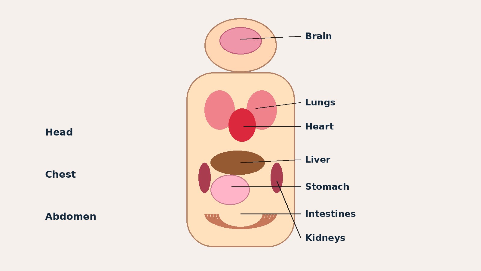
- The brain coordinates sensation, movement, thought, memory, and many automatic body processes.
- The heart pumps blood around the body, while the lungs move oxygen into the blood and carbon dioxide out of it.
- The stomach and intestines break food down and absorb nutrients, with help from the liver, gallbladder, and pancreas.
- The kidneys and bladder handle urine: the kidneys filter blood and form urine, and the bladder stores urine until it leaves the body.
- The skin covers and protects the body, controls temperature, and senses touch, pressure, temperature, and pain.
Introduction
A diagram of the human body can look crowded. Organs overlap and share small spaces, so it is easy to mix up neighbors such as the liver and the gallbladder, or the pancreas and the spleen. This guide untangles that picture organ by organ.
It is an organs overview, not a tour of body systems. A system name is mentioned only where it helps explain an organ’s job; the focus stays on the organ itself: where it sits, what it does, and how it fits beside its neighbors.
What Is an Organ?
Cells are the smallest independently functioning units of a living organism. A tissue is a group of many similar cells working together for a specific function. An organ is an anatomically distinct structure composed of two or more tissue types, each performing one or more specific physiological functions. A group of organs working together for major functions is an organ system, and all those levels together make up the organism.
That definition explains why a bone counts as more than a hard stick, why skin counts as more than a wrapper, and why a small gland such as the thyroid counts beside the liver, because an organ is defined by organized structure and job, not size alone.
Counting organs is less simple than it sounds. Textbooks group and name structures in slightly different ways, and some structures count as one organ in one list and several parts in another. A list of major organs is therefore a learning map, not a final census. The organs here are the ones most anatomy overviews include, grouped by where they sit so they are easier to picture.
A hollow organ such as the stomach or bladder can stretch and hold material. A solid organ such as the liver or kidney is packed with working tissue and blood vessels. A muscular organ such as the heart is built to contract again and again. When you learn an organ, learn three things together: where it is, what it does, and what structure lets it do that job.
Organs at a Glance
| Organ | Main location | Main job |
|---|---|---|
| Brain | Inside the skull | Coordinates thought, movement, and regulation |
| Spinal cord | Inside the vertebral column | Carries signals between brain and body |
| Thyroid gland | Front of the neck | Releases hormones for metabolism |
| Heart | Chest, between the lungs | Pumps blood around the body |
| Lungs | Chest, one on each side of the heart | Exchange oxygen and carbon dioxide |
| Stomach | Upper left abdomen | Stores food, begins breaking it down |
| Small intestine | Central and lower abdomen | Digests food, absorbs nutrients |
| Large intestine | Abdomen, framing the small intestine | Absorbs water, forms solid waste |
| Liver | Upper right abdomen | Processes nutrients, makes bile |
| Gallbladder | Beneath the liver | Stores and concentrates bile |
| Pancreas | Behind the stomach | Makes enzymes and hormones |
| Spleen | Upper left abdomen, behind the stomach | Filters blood, supports immune defense |
| Kidneys | Back of the abdomen, by the spine | Filter blood, form urine |
| Bladder | Pelvis | Stores urine |
| Skin | Covering the whole body | Protects, senses, regulates temperature |
How to Read Where an Organ Sits
Anterior means toward the front of the body, posterior toward the back, superior above, inferior below, medial toward the midline, and lateral toward the side. Right and left in anatomy are the person’s right and left, not the viewer’s, which matters when the liver appears on your left side of a front-view diagram.
The body also has cavities, spaces that hold organs: the cranial cavity inside the skull holds the brain, the spinal cavity inside the vertebral column holds the spinal cord, the thoracic (chest) cavity holds the heart and lungs, the abdominal cavity holds most digestive organs, and the pelvic cavity holds the bladder and reproductive organs. The diaphragm, a sheet of muscle below the lungs, separates the chest from the abdomen. Kidneys sit at the back of the abdomen, behind the main abdominal space.
Depth matters too: an organ described as behind the stomach is deeper in the body, not beside it on a flat diagram. Neighbors also hint at jobs: the liver sits close to the gallbladder because bile made there is stored there, and the pancreas sits behind the stomach because its juices need a short route into the small intestine.
Head and Neck Organs
Brain
The brain sits inside the cranial cavity, protected by the skull. It receives sensory information, processes it, and sends instructions for movement, speech, memory, emotion, and thought. It also helps regulate automatic processes such as breathing and heart rate, through the spinal cord and nerves. Different regions handle different tasks, so a change in one region does not affect every function alike.
Spinal cord
The spinal cord runs through the vertebral column from the base of the brain toward the lower back. It carries sensory signals up toward the brain and motor signals down toward muscles and glands. It also carries out some rapid, local responses without waiting for a full brain decision. Think of it as the main signal highway, with nerves branching to the arms, trunk, and legs.
Eyes, ears, nose, and tongue
The sense organs of the head turn outside information into signals the brain can use. The eyes detect light and help form vision. The ears detect sound, and the inner ear also helps with balance. The nose detects odors and warms and filters air on its way toward the lungs. The tongue detects taste and helps move food during chewing and swallowing. They are small beside the liver or lungs, but organs in the same structural sense.
Thyroid and nearby neck glands
The thyroid gland sits at the front of the neck, below the voice box, and wraps partly around the upper windpipe. It releases hormones that help set the pace of metabolism, the chemical processes that turn food into energy and building material. The small parathyroid glands sit on the back of the thyroid and help regulate blood calcium. The pituitary gland at the base of the brain releases hormones that influence other glands, including the thyroid. Textbooks often call it a master control gland, but that nickname should not be taken literally: it works as part of a feedback loop, not as a lone commander.
Chest Organs
Heart
The heart sits in the thoracic cavity, between the lungs and slightly to the left of the midline. It is a muscular, hollow organ with four chambers: two upper and two lower. One route sends blood to the lungs to pick up oxygen and release carbon dioxide; the other sends oxygen-rich blood out to the body and brings blood back for another pass. Valves keep blood moving in one direction, and the heart muscle is built for steady, repeated contraction.
Lungs
The lungs sit in the thoracic cavity, one on each side of the heart, protected by the rib cage. Air travels down the windpipe, called the trachea, which divides into airways called bronchi, then into smaller tubes ending in tiny air sacs called alveoli. In the alveoli, oxygen moves from air into the blood, and carbon dioxide moves from blood into air to be breathed out. The right lung is generally broader, with three lobes, while the left lung is narrower, with room for the heart, and has two lobes.
Esophagus and trachea: two tubes, two jobs
The trachea carries air toward the lungs. The esophagus carries food and drink from the mouth toward the stomach, and sits behind the trachea for much of its course. Food does not enter the lungs during normal swallowing: a coordinated swallowing action directs food into the esophagus and keeps the airway clear. Remembering which tube does which job prevents many diagram mistakes.
Thymus
The thymus sits in the upper chest, behind the breastbone and in front of the heart. It plays a role in developing immune cells, especially in early life. It is largest in childhood and becomes smaller with age, as fatty tissue gradually replaces much of its working tissue.
Abdomen Organs for Digestion
Digestion is a relay. Food enters through the mouth, where teeth break it into smaller pieces and saliva begins to moisten it, then travels through the esophagus into the stomach, and each later organ changes it in a different way.
Stomach
The stomach sits in the upper left abdomen, below the diaphragm. It is a muscular, stretchy, pouch-shaped organ. It stores food, mixes it with acid and enzymes, and slowly releases the partly broken-down mixture into the small intestine. Its wall churns contents, helping break food into smaller particles. The stomach begins digestion, it does not finish it.
Small intestine
The small intestine is a long, coiled tube in the central and lower abdomen. Despite its name, it is small in width, not length, and longer than the large intestine. Digestive enzymes from the pancreas and bile from the liver and gallbladder help break food down further, and the inner wall absorbs nutrients into the blood. Its inner surface is folded and covered with tiny projections that increase the area available for absorption. If you remember one job here, make it absorption.
Large intestine
The large intestine frames the small intestine in the abdomen. It is wider and shorter than the small intestine. It absorbs water and salts from what remains after the small intestine has done its work, and forms solid waste for removal. It also hosts a large community of microbes breaking down some remaining material. The colon is the longest part of the large intestine, ending at the rectum. Its job is finishing and forming, not starting digestion.
Liver
The liver sits in the upper right abdomen, just below the diaphragm. It is the largest internal organ. It processes nutrients from the digestive tract, stores energy and vitamins for later use, makes proteins needed in the blood, and helps handle substances needing change or removal. It also makes bile, a fluid that helps digest fats. Textbooks often call it a metabolic hub, but that is a summary, not a single function.
Gallbladder
The gallbladder is a small, pouch-shaped organ tucked beneath the liver. It stores bile made by the liver and concentrates it, then releases it into the small intestine when fatty food arrives. A common confusion is to credit the gallbladder with making bile. It does not make bile, it stores and releases it.
Pancreas
The pancreas sits behind the stomach, stretching across the upper abdomen from right to left. It has two kinds of work, so it appears in both digestive and hormone discussions. As a digestive organ, it releases enzymes into the small intestine that help break down carbohydrates, proteins, and fats. As a hormone organ, it releases hormones into the blood, including insulin and glucagon, which help regulate blood sugar. Its position behind the stomach explains why front-view diagrams often hide it.
Spleen
The spleen sits in the upper left abdomen, behind the stomach and near the left kidney. It filters blood, removes worn-out red blood cells, and supports immune defense. It sits close to the stomach and pancreas on a diagram, but it is not part of the food relay: no food passes through it.
Abdomen Organs for Filtering and Balance
Kidneys
The kidneys are a pair of bean-shaped organs in the back of the abdomen, one on each side of the spine, roughly at the level of the lower ribs. Each kidney filters blood, removes waste products and excess water and salts, and forms urine. They also help regulate fluid balance, salt balance, and blood pressure, and help produce a hormone that supports red blood cell production. Urine leaves each kidney through a tube called a ureter, which carries it down to the bladder. The kidneys filter; the bladder stores. Keeping those roles separate avoids a very common mix-up.
Adrenal glands
One adrenal gland sits on top of each kidney. Despite the shared address, they are separate organs with their own jobs. They release hormones that help the body respond to stress and regulate salt balance, blood pressure, and metabolism. Adrenaline is the best-known adrenal hormone, but it is not the only one, and the outer and inner parts of each gland release different hormones. Location is the link to the kidney here, not function.
Bladder
The bladder sits in the pelvic cavity, in the lower center of the abdomen. It is a hollow, muscular organ that stores urine until it is released through the urethra. Its wall stretches as it fills and contracts when it empties. Like the stomach, it is a storage organ with a muscular wall, but its contents and route out differ entirely.
Pelvis and Reproductive Organs
Reproductive organs differ between female and male bodies, and a general overview names them without becoming a reproduction guide. In female anatomy, the main internal reproductive organs are the ovaries, the fallopian tubes, the uterus, and the vagina. The ovaries produce eggs and sex hormones. The fallopian tubes carry eggs toward the uterus. The uterus is a muscular organ where a pregnancy develops. In male anatomy, the main reproductive organs include the testes, which produce sperm and sex hormones, and a set of ducts and glands that store, nourish, and transport sperm, including the prostate gland.
The shared pattern is the useful point: a reproductive organ makes reproductive cells, makes hormones, or moves cells toward fertilization. Details belong in a dedicated reproduction guide; this overview keeps the list at names, places, and main jobs.
Glands as Organs
A gland is an organ or part of an organ that releases a substance, either into a duct or directly into the blood. Digestive glands release enzymes or fluids into tubes. Hormone glands, also called endocrine glands, release hormones into the blood, where they travel to target organs.
The main hormone glands here are the pituitary gland at the base of the brain, the thyroid and parathyroid glands in the neck, the adrenal glands on the kidneys, the hormone-producing part of the pancreas, and the ovaries or testes. The salivary glands in the mouth, which release saliva, are digestive glands, not hormone glands. Sorting glands by what they release and where it goes beats memorizing a single list, because the pancreas can appear in both groups for different reasons.
Skin, Bones, and Muscles: Borderline Cases
Some lists include skin, bones, and muscles as organs; others treat them as tissues or systems. Both views have a reason, and learners should know why the disagreement exists.
Skin is widely treated as an organ. It protects against injury and microbes, senses touch, pressure, temperature, and pain, controls body temperature through sweating and blood flow near the surface, and produces vitamin D when exposed to sunlight. Its layers of different tissues fit the organ definition.
A single bone is also an organ in the structural sense: it contains bone tissue, cartilage, blood vessels, and nerves, and it supports, protects, and helps move the body. Bone tissue also stores minerals and, in the marrow of some bones, produces blood cells. Skeletal muscles are organs in the same sense: each muscle is a distinct structure of muscle tissue, connective tissue, blood vessels, and nerves, built to contract. Textbooks sometimes discuss bones and muscles as systems rather than listing every one as an organ, because there are so many. Skin, a bone, and a skeletal muscle each meet the organ definition, but overviews group them as systems when a full list would be unmanageably long.
How Organs Work Together Without Becoming a Systems Lesson
Organs rarely work alone, and three short examples show the pattern without making this a systems article. Breathing needs lungs, a windpipe, a diaphragm muscle, and a heart to carry oxygen onward. Eating needs the mouth, stomach, small intestine, liver, gallbladder, and pancreas, each changing food or its surroundings in turn. Filtering blood needs kidneys to form urine, ureters to carry it, and a bladder to store it. In each case, the organs stay distinct, but the result belongs to the chain.
This is also why lists sometimes disagree about which group an organ belongs to: the pancreas belongs in a digestive list and a hormone list, the spleen with blood filtering and immune defense, and the liver with digestion and chemical processing. An organ can serve more than one chain, and a good organs guide notes the second job instead of forcing it into a single box.
If you have a health concern, speak with a qualified health professional.
Common Organ Mix-Ups
Liver and gallbladder
The liver makes bile. The gallbladder stores and concentrates bile, releasing it when needed. If a diagram labels the small pouch as the bile maker, the label is wrong.
Pancreas and spleen
Both sit near the stomach in the upper left abdomen, but their jobs differ. The pancreas releases digestive enzymes and hormones. The spleen filters blood and supports immune defense. Food does not pass through either organ.
Kidneys and bladder
The kidneys filter blood and form urine. The bladder stores urine. The ureters connect them. Naming the bladder as a filter, or the kidneys as storage, reverses the relay.
Small intestine and large intestine
Small refers to width, not length. The small intestine is longer and narrower and absorbs most nutrients. The large intestine is wider and shorter and absorbs water and forms waste.
Brain and spinal cord
The brain coordinates and integrates. The spinal cord carries signals and handles some local responses. Together they form the central nervous system, but they are separate organs with separate places and jobs.
Heart and lungs
The heart pumps blood. The lungs exchange gases with blood. They sit side by side in the chest, depending on each other, but pumping and gas exchange are different jobs.
How to Study an Organs Diagram
Start with the big landmarks: skull, rib cage, diaphragm, and pelvis. Place the brain in the skull, the heart and lungs in the chest, the liver on the abdomen’s upper right, the stomach on the upper left, and the bladder in the pelvis. Then add the hidden organs: pancreas behind the stomach, kidneys at the back of the abdomen, and spleen behind the stomach on the left. Finally add the glands: thyroid in the neck, adrenals on the kidneys, and pituitary at the base of the brain.
Read labels in pairs: organ and job, organ and neighbor, organ and route. For a tube organ, follow its route. For a gland, ask what it releases and where it goes.
Conclusion
An organ is a distinct structure of two or more tissues doing a specific job, and the major organs repay learning in place. The brain coordinates, the heart pumps, the lungs exchange gases, the stomach and intestines digest and absorb, the liver processes and makes bile, the pancreas adds enzymes and hormones, the kidneys filter and form urine, the bladder stores urine, and the skin protects and senses. Keep place, structure, and job together for each organ, watch for organs with two jobs, and the crowded torso diagram becomes readable.
Frequently Asked Questions
What is an organ in simple terms?
An organ is a distinct body structure made from two or more tissue types working together for one or more specific jobs. Examples include the heart, lungs, liver, stomach, kidneys, brain, and skin. Tissues are groups of similar cells; organs are built from tissues; groups of organs working together form organ systems.
How many major organs are in the human body?
There is no single agreed count, because textbooks group structures differently. Some lists count around seventy or more organs, while shorter learning lists name twenty to thirty major organs. The difference is grouping: for example, whether each bone or muscle is counted separately.
What is the largest organ in the human body?
Skin is commonly listed as the body’s largest organ by area, covering the whole body, while the liver is commonly listed as the largest internal organ. Largest by outside area and largest inside the body are different measures, so both answers appear in overviews.
What do the liver, gallbladder, and pancreas each do?
The liver processes nutrients, makes bile, and carries out many chemical tasks. The gallbladder stores bile made by the liver and releases it into the small intestine. The pancreas releases digestive enzymes into the small intestine and hormones, including insulin, into the blood.
Why are there two lungs and two kidneys?
Paired organs provide capacity and some backup, and their positions reflect the space around them. Two lungs fill the two sides of the chest around the heart, and two kidneys sit on either side of the spine at the back of the abdomen. Paired does not mean identical: the left lung is narrower to make room for the heart.
Where is the pancreas, and why is it hard to find on diagrams?
The pancreas sits behind the stomach in the upper abdomen, stretching from right to left. Front-view diagrams often hide it behind the stomach and liver, so learners miss it or mistake it for part of the stomach. Looking behind the stomach, rather than beside it, makes it easier to spot.
What does the spleen do?
The spleen filters blood, removes worn-out red blood cells, and supports immune defense. It sits in the upper left abdomen, behind the stomach. It is not part of the food relay, though it sits close to digestive organs on a diagram.
Is skin really an organ?
Yes, in the structural sense used in anatomy. Skin is made from more than one tissue type, covers the body as a distinct structure, and performs specific jobs: protection, sensation, temperature regulation, and vitamin D production with sunlight exposure. Overviews that focus on internal organs sometimes omit it for space, not because it fails the definition.
What is the difference between an organ and an organ system?
An organ is one distinct structure with one or more specific jobs, such as the heart pumping blood. An organ system is a group of organs working together for major functions or physiological needs, such as the organs digesting food together. This guide focuses on organs; a systems guide would focus on how each group works as a whole.
Sources & references
- OpenStax, Anatomy and Physiology 2e, Section 1.2 Structural Organization of the Human Body: organ, tissue, and organ system definitions and levels of organization. https://openstax.org/books/anatomy-and-physiology-2e/pages/1-2-structural-organization-of-the-human-body
- OpenStax, Anatomy and Physiology 2e, Section 19.1 Heart Anatomy: heart position, chambers, and circulation routes. https://openstax.org/books/anatomy-and-physiology-2e/pages/19-1-heart-anatomy
- OpenStax, Anatomy and Physiology 2e, Section 22.1 Organs and Structures of the Respiratory System: trachea, esophagus position, and gas exchange at the alveoli. https://openstax.org/books/anatomy-and-physiology-2e/pages/22-1-organs-and-structures-of-the-respiratory-system
- OpenStax, Anatomy and Physiology 2e, Section 22.2 The Lungs: lung lobes and room for the heart. https://openstax.org/books/anatomy-and-physiology-2e/pages/22-2-the-lungs
- OpenStax, Anatomy and Physiology 2e, Section 23.5 Accessory Organs in Digestion: liver, pancreas, and gallbladder roles in digestion. https://open.oregonstate.education/aandp/chapter/23-5-accessory-organs-in-digestion-the-liver-pancreas-and-gallbladder/
- Cleveland Clinic, Organs in the Body: Definition & Anatomy: organ listings, largest organ by area, largest internal organ, and organ-count variation. https://my.clevelandclinic.org/health/articles/organs-in-the-body


