Short answer
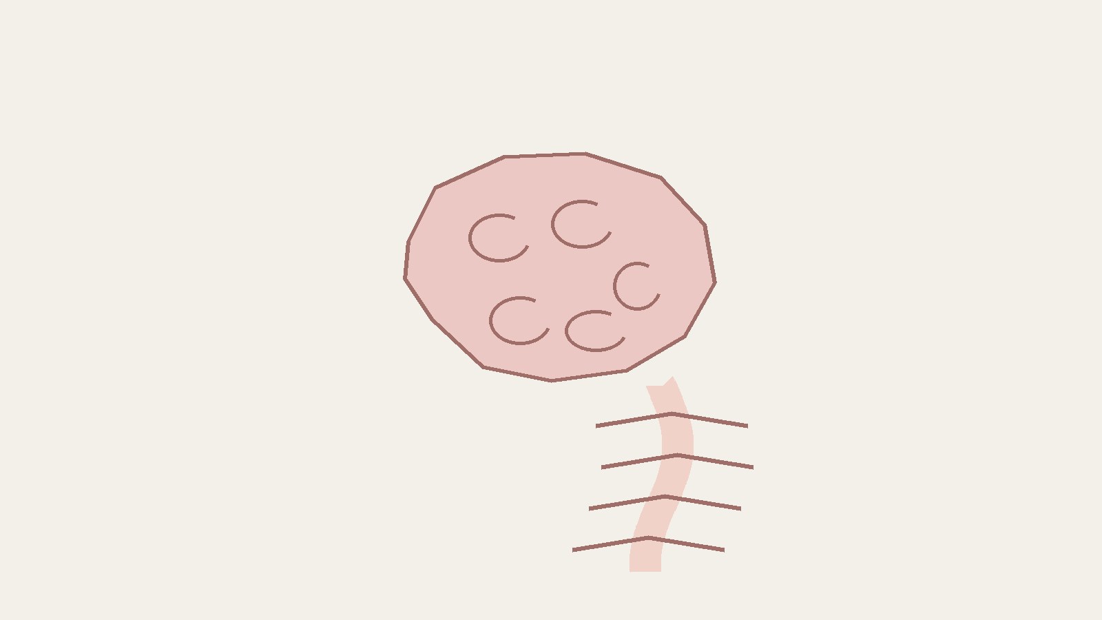
- The nervous system is the network of nerve cells and supporting cells that carries signals around the body, processes those signals, and produces responses. In humans it is made up of the brain, the spinal cord, and the nerves that connect them to the rest of the body. It detects changes inside and outside the body, decides what those changes mean, and then sends instructions to muscles and glands. Its work can be grouped into three overlapping jobs. First, it senses: receptors detect stimuli such as light, sound, temperature, pressure, and changes inside the body. Second, it integrates: the brain and spinal cord process that sensory information, compare it with other information and with memory, and form a response. Third, it acts: signals travel outward to muscles, which contract, or to glands, which release secretions. Together, these three jobs let the body move, breathe, see, think, learn, and remember, and they help keep internal conditions stable. Anatomically, the nervous system has two main parts. The central nervous system, usually shortened to CNS, is the brain and spinal cord. The peripheral nervous system, usually shortened to PNS, is made up of the nerves that branch off from the brain and spinal cord and extend to all parts of the body. The basic unit of the system is the neuron, a nerve cell. Bundles of axons, the long extensions of neurons, form nerves. Supporting cells called glia help neurons work properly, protect them, and make myelin, the insulation that helps nerve impulses travel.
What the nervous system is, in plain terms
A useful way to picture the nervous system is as the body’s communication and control network. It links other systems together by moving information, not substances, from place to place. When you step on a sharp stone, you do not have to think through each step of what happens next. Receptors in your foot detect pressure, a signal travels to the spinal cord and brain, and a signal returns to the muscles of your leg. You lift your foot, and you become aware of pain, almost at the same time. That whole loop is the nervous system at work.
The United States National Cancer Institute training material describes the nervous system as the major controlling, regulatory, and communicating system in the body, and as the center of all mental activity including thought, learning, and memory. Thought, learning, memory, emotion, and automatic processes such as heartbeat and breathing all depend on the same network.
The nervous system does not work alone. Together with the endocrine system, the system of glands that releases hormones into the blood, it regulates and maintains homeostasis. Homeostasis is the maintenance of relatively stable internal conditions, such as body temperature and the balance of salts and fluids, even when conditions outside the body change. The two systems work on different timescales. Nervous signals are fast and targeted, arriving at a specific muscle or gland within a fraction of a second. Hormones travel in the blood and act more slowly and more widely. In many everyday responses, both systems act together.
The two main parts: central and peripheral
Anatomists divide the nervous system first by location. The central nervous system is the brain and spinal cord. The peripheral nervous system is everything else: the nerves and clusters of nerve cell bodies, called ganglia, that lie outside the brain and spinal cord. This is an anatomical division, a division by where structures sit, not a division by what they do. Many functions cross the boundary. A single neuron can have its cell body in the central nervous system and its axon extending far into the peripheral nervous system.
The central nervous system is protected by bone and by membranes. The brain sits inside the cranium, formed by the skull. The spinal cord runs inside the vertebral canal, formed by the stacked bones of the spine. Three layers of membranes, called meninges, surround both. The outer layer is the dura mater, the middle layer is the arachnoid, and the innermost layer is the pia mater.
The peripheral nervous system connects the central nervous system to the rest of the body. Nerves that exit from the brain are called cranial nerves. Nerves that exit from the spinal cord are called spinal nerves. OpenStax material on the spinal cord records 31 pairs of spinal nerves, organized by the region of the spine from which they emerge: cervical nerves from the neck, thoracic nerves from the upper back, lumbar nerves from the lower back, and sacral nerves from the pelvis. Each spinal nerve carries both sensory fibers, bringing information toward the central nervous system, and motor fibers, carrying instructions away from it.
| Division | Main structures | Main job |
|---|---|---|
| Central nervous system (CNS) | Brain and spinal cord | Process information, integrate signals, form responses, store memories |
| Peripheral nervous system (PNS) | Cranial nerves, spinal nerves, ganglia | Carry sensory information to the CNS and carry motor instructions from the CNS to muscles and glands |
The brain is not the whole nervous system. On its own it would be isolated: without the spinal cord and peripheral nerves, it could neither receive information from the body nor send instructions back to it. The spinal cord is not a passive cable either. It conducts signals in both directions and contains circuits that process information locally, which is why some reflexes can happen without direct involvement of the brain.
The cells of the nervous system: neurons and glia
The basic unit of the nervous system is a nerve cell, or neuron. The human brain contains about 100 billion neurons, according to the Eunice Kennedy Shriver National Institute of Child Health and Human Development. That figure is an estimate, and it gives a sense of scale rather than an exact count. What matters more than the total is how neurons are organized. Neurons form networks, and the pattern of connections in a network is what allows different parts of the brain to talk to each other and work together to control body functions, emotions, thinking, and behavior.
A typical neuron has three main parts. The cell body contains the nucleus and most of the cell machinery. Dendrites are shorter extensions that receive signals from other neurons and pass them toward the cell body. The axon is a single longer extension that carries the signal away from the cell body toward other neurons, muscles, or glands. Some axons are very short, connecting neighboring cells. Others are very long. The axons that run from the spinal cord to the muscles of the foot, for example, can be about a meter long in an adult.
Neurons are not the only cells in the nervous system. The other major class is glia, also called neuroglia or glial cells, pronounced GLEE uh. Glia were long described only as support cells, from a Greek word for glue. That picture is incomplete. Glia help support and hold neurons in place, protect neurons, repair neurons, trim out dead neurons, and regulate neurotransmitters, the chemical messengers described below.
One of the most important glial jobs is making myelin. Myelin is a fatty insulation that wraps around many axons and helps nerve impulses travel faster and more efficiently. In the central nervous system, myelin is produced by cells called oligodendrocytes. In the peripheral nervous system, it is produced by Schwann cells. Other glial types have their own roles. Astrocytes, named for their star shape, are abundant in the central nervous system and help regulate the chemical environment around neurons, take up or break down some neurotransmitters, and help form the blood brain barrier, the boundary that separates the circulatory system from the brain. Microglia protect the nervous system against infection. A nervous system made only of neurons, without glia, would not function for long.
Neurons come in different functional types. Sensory neurons detect stimuli and send messages toward the central nervous system. Motor neurons transmit messages to muscles or glands. Interneurons, found mainly in the central nervous system, connect sensory and motor neurons and form the circuits that process information.
How a signal travels: impulses, synapses, and neurotransmitters
Nervous communication uses two kinds of signal in sequence: electrical and chemical. When a neuron sends a message, it first sends an electrical signal down the length of its axon. That traveling electrical signal is called a nerve impulse, or action potential. It begins when a stimulus changes the electrical state of the neuron membrane, the boundary of the cell. If the change is large enough, it triggers a wave of electrical change that travels along the axon to its end. Myelin speeds this process, because the impulse can jump between gaps in the myelin sheath rather than traveling continuously along the whole axon.
At the end of the axon, the signal must cross to the next cell, and here it changes form. The electrical signal changes to a chemical signal. The axon releases chemical messengers called neurotransmitters into the synapse, the small space between the end of one axon and the tip of a dendrite from another neuron. The neurotransmitters cross the synapse and bind to the neighboring dendrite, which converts the chemical signal back into an electrical signal. The electrical signal then travels through the next neuron, and the same conversion process repeats at each synapse along the chain.
The synapse is therefore more than a gap. It is a control point. Whether the next neuron fires depends on how much neurotransmitter is released, which type it is, and what other signals the receiving neuron is getting at the same time. Some neurotransmitters excite the next neuron, others inhibit it, and a single neuron may receive thousands of inputs. Its response depends on the balance among them. That balancing act is the physical basis of integration.
Three jobs: sensory function, integration, and motor function
The National Cancer Institute training material groups the activities of the nervous system into three general, overlapping functions: sensory, integrative, and motor. OpenStax Anatomy and Physiology uses the same three terms, calling them sensation, integration, and response. Keeping the three jobs separate helps a reader follow any example, from catching a ball to reading this sentence.
Sensation is receiving information about the environment and translating it into the electrical signals of nervous tissue. Millions of sensory receptors detect changes, called stimuli, inside and outside the body. From the external environment they monitor temperature, light, and sound. Inside the body they detect variations in pressure, pH, carbon dioxide concentration, and the levels of various electrolytes, as well as the position of limbs and the stretch of muscles and organs. Sensory information travels toward the central nervous system through the peripheral nerves in the division known as the afferent, or sensory, branch. When information arises from receptors in the skin, skeletal muscles, or joints, it is transmitted by somatic sensory neurons. When it arises from receptors in blood vessels or internal organs, it is transmitted by visceral sensory neurons.
Integration is the processing step. In the central nervous system, information from one stimulus is compared with, or integrated with, information from other stimuli or memories of previous stimuli. Sensory input is brought together to create sensations, to produce thoughts, or to add to memory. Decisions are made each moment based on sensory input. Integration is where context enters. The same pattern of light on the retina can be read as a familiar face, a stranger, or a photograph, depending on memory and on other information arriving at the same time.
Motor function is the response. Based on sensory input and integration, the nervous system sends signals to muscles, causing them to contract, or to glands, causing them to produce secretions. Muscles and glands are called effectors, because they cause an effect in response to directions from the nervous system. The motor, or efferent, branch of the peripheral nervous system carries signals away from the central nervous system to the effector organs. When the effector is a skeletal muscle, the neuron carrying the information is called a somatic motor neuron. When the effector is cardiac muscle, smooth muscle, or glandular tissue, it is called an autonomic motor neuron.
For example, when you read a word, light reaches receptors in the retina, sensory neurons carry the pattern to the brain, the brain integrates it with knowledge of language, and motor neurons move the eyes to the next word.
The brain: the main processing center
The brain is the large organ of the central nervous system contained within the cranium and continuous with the spinal cord. It is where most integration happens, and it is the center of thought, learning, memory, language, emotion, and conscious perception. It also controls many processes that run without conscious attention, such as breathing and the regulation of body temperature, through structures in its lower parts.
A broad map of the brain divides it into three regions: the cerebrum, the cerebellum, and the brainstem. The cerebrum is the largest part. Its outer layer, the cerebral cortex, is involved in conscious perception, thought, language, memory, and voluntary movement. Different areas of the cortex specialize in different tasks, such as vision, hearing, touch, movement, and planning, but they work as connected networks rather than as isolated modules. The cerebellum sits at the back of the brain, below the cerebrum. It is best known for coordination, balance, and the fine tuning of movement. The brainstem connects the brain to the spinal cord. It contains structures that regulate vital automatic functions, including breathing, heart rate, and blood pressure, and it serves as a pathway for signals traveling between the brain and the rest of the body.
The brain has left and right halves, called hemispheres, connected by a large bundle of fibers. It is also organized into lobes, named for the bones of the skull that cover them: frontal, parietal, temporal, and occipital. The frontal lobe, at the front, is heavily involved in planning, decision making, and voluntary movement. The parietal lobe, behind it, helps process touch and spatial information. The temporal lobe, at the side, helps process hearing and memory. The occipital lobe, at the back, is primarily involved in vision.
The spinal cord and reflexes
The spinal cord is the extension of nervous tissue within the vertebral column, continuous with the brain at its upper end. It is contained within the vertebral canal and connected with the periphery through the spinal nerves. Its two main roles are conduction and local processing. As a conduction pathway, it carries sensory information upward to the brain on ascending tracts and motor instructions downward from the brain on descending tracts. As a processing center, it contains circuits that can produce responses on their own.
The reflex is the clearest example of spinal processing. A reflex is a fast, automatic response to a stimulus that does not require conscious decision. The classic example is touching a hot surface and pulling the hand away. Sensory receptors in the skin detect extreme temperature. The signal travels along a sensory neuron into the spinal cord. There it connects, directly or through an interneuron, to a motor neuron, which carries a signal back out to the muscles of the arm, causing them to contract and withdraw the hand. At the same time, a signal travels upward to the brain, so you become aware of the heat and the pain. Awareness follows the withdrawal rather than causing it, which is why the hand can move before you have consciously decided to move it.
Other reflexes, such as blinking and the knee jerk, follow the same design. Voluntary movement, such as choosing to pick up a cup, involves processing in the brain before motor signals are sent.
The peripheral nervous system in more detail
Beyond the central versus peripheral split, the peripheral nervous system is divided by function into somatic and autonomic parts. A single peripheral nerve can contain both somatic and autonomic fibers, just as it contains both sensory and motor fibers.
The somatic nervous system is the part of the peripheral nervous system associated with voluntary control of skeletal muscles and with conscious perception of the environment. Somatic sensory neurons bring information from the skin, muscles, and joints to the central nervous system, giving rise to sensations such as touch, pressure, temperature, pain, and the sense of limb position. Somatic motor neurons carry instructions from the central nervous system to skeletal muscles, producing voluntary movement and skeletal muscle reflexes.
The autonomic nervous system regulates involuntary responses. It controls cardiac muscle, smooth muscle, and glands, and therefore governs processes such as heart rate, blood pressure, digestion, sweating, pupil size, and the emptying of the bladder. The autonomic nervous system is itself divided into three parts: sympathetic, parasympathetic, and enteric.
The sympathetic nervous system is commonly known as the fight or flight system. It is activated when the body is under actual or perceived stress, to ready the body response to a potential threat. Activation produces a coordinated set of changes: increased heart rate and force of heart contraction, increased blood pressure, shunting of blood to skeletal muscle, widening of the airways, dilation of the pupils, decreased salivation and digestion, release of glucose into the blood, and release of the hormones epinephrine and norepinephrine. The parasympathetic nervous system acts in the opposite direction. It is activated when the body is in a relaxed state, and it promotes rest, digestion, and recovery. It slows the heart, constricts the pupils, stimulates salivation and digestive activity, and supports the processes that rebuild energy stores after activity.
The sympathetic and parasympathetic systems are often described as opposing each other, but in most situations both are active, and the balance between them sets the state of the organs they control. The heart, for example, receives both kinds of input at all times.
The enteric nervous system is the third autonomic division. It is a large network of neurons embedded in the walls of the gastrointestinal tract, from the esophagus to the anus, and it controls the movement and secretions of the gut. It can operate with a high degree of independence from the brain and spinal cord. That independence should not be overstated: the enteric nervous system does not think or form memories in the way the brain does, but its size and independence reflect how complex the control of digestion needs to be, with its own sensory neurons, interneurons, and motor neurons forming complete local circuits.
Senses: how the outside world becomes a signal
Each sense depends on specialized receptors that convert one form of energy into a nerve signal, a process called transduction. Photoreceptors in the retina convert light into signals. Hair cells in the inner ear convert sound vibrations, and also head movement, into signals. Chemoreceptors in the nose and tongue convert chemical molecules into signals for smell and taste. Mechanoreceptors in the skin, muscles, and joints convert pressure, vibration, stretch, and touch into signals. Thermoreceptors convert temperature changes into signals.
Proprioceptors in muscles, tendons, and joints report the position of the limbs, which is how you can touch your nose with your eyes closed. These internal senses are essential to homeostasis, because they tell the central nervous system what adjustments are needed.
Common misunderstandings
First, the brain is not the whole nervous system. It is the most complex part, but the spinal cord, the peripheral nerves, and the enteric network are also parts of the system, and they carry out work the brain cannot do alone.
Second, nerves are not single cells. A nerve is a bundle of axons from many different neurons, wrapped in connective tissue. A single neuron is a cell. A nerve is a cable made of many cell fibers together.
Third, the left brain and right brain are not separate minds. The two hemispheres specialize to some degree, but they are connected by a large bundle of fibers and work together on almost every task.
Fourth, the nervous system and the endocrine system are not rivals. They are partners with different speeds and ranges, and many everyday responses, from reacting to a loud noise to waking in the morning, involve both.
FAQ
What is the nervous system in simple terms? It is the body’s signal network. It is made up of the brain, spinal cord, and nerves. It senses changes, processes the information, and sends instructions to muscles and glands.
What are the two main parts of the nervous system? The central nervous system, which is the brain and spinal cord, and the peripheral nervous system, which is the nerves that branch off from the brain and spinal cord and extend to the rest of the body.
What is the difference between the central and peripheral nervous systems? The difference is anatomical, based on location. The central nervous system is the brain and spinal cord, protected inside the skull and spine. The peripheral nervous system is everything outside those structures, carrying signals to and from the central nervous system.
What is a neuron? A neuron is a nerve cell, the basic unit of the nervous system. It has a cell body, dendrites that receive signals, and an axon that carries signals away.
How do neurons communicate? A neuron sends an electrical signal, called a nerve impulse, along its axon. At the end of the axon, the signal becomes chemical. The axon releases neurotransmitters into the synapse, the gap to the next cell, and those chemicals trigger a new electrical signal in the receiving cell.
What is the difference between a neuron and a nerve? A neuron is a single cell. A nerve is a bundle of axons from many neurons, wrapped in connective tissue. Nerves are the cables of the peripheral nervous system.
What are the three functions of the nervous system? Sensation, integration, and motor function. Sensation gathers information through receptors. Integration processes that information in the brain and spinal cord. Motor function sends responses to muscles and glands.
What is the somatic nervous system? It is the functional division of the peripheral nervous system that handles conscious perception and voluntary movement of skeletal muscles. Somatic sensory neurons bring information from the skin, muscles, and joints to the central nervous system, and somatic motor neurons carry instructions back to skeletal muscles.
What is the autonomic nervous system? It regulates involuntary processes, including heart rate, digestion, sweating, and pupil size. It is divided into sympathetic, parasympathetic, and enteric parts.
What is the difference between the sympathetic and parasympathetic nervous systems? The sympathetic system prepares the body for action under stress, increasing heart rate and blood pressure. The parasympathetic system promotes rest and recovery, slowing the heart and supporting digestion. In most situations both are active, and the balance between them sets the state of the organs they control.
What is a reflex? A reflex is a fast, automatic response to a stimulus, produced by a short circuit through the spinal cord or brainstem without requiring a conscious decision. Withdrawing the hand from a hot surface is a classic example.
How many neurons are in the human brain? The Eunice Kennedy Shriver National Institute of Child Health and Human Development states that the human brain contains about 100 billion neurons. That is an estimate that gives a sense of scale, not an exact count.
What is myelin and why does it matter? Myelin is fatty insulation around many axons that helps nerve impulses travel faster and more efficiently. In the central nervous system it is made by oligodendrocytes, and in the peripheral nervous system by Schwann cells.
What is the enteric nervous system? It is the network of neurons in the walls of the gastrointestinal tract that controls gut movement and secretions. It can operate with a high degree of independence, but it is part of the autonomic nervous system, not a separate brain that thinks.
Keep reading: How to Draw a Brain
Sources & references
- Eunice Kennedy Shriver National Institute of Child Health and Human Development, National Institutes of Health. What are the parts of the nervous system? Accessed 27 October 2026. https://www.nichd.nih.gov/health/topics/neuro/conditioninfo/Pages/parts.aspx
- National Cancer Institute, National Institutes of Health, SEER Training Modules. Introduction to the Nervous System. Accessed 27 October 2026. https://training.seer.cancer.gov/anatomy/nervous/
- OpenStax, Anatomy and Physiology 2e, Chapter 12, Structure and Function of the Nervous System, as republished by Oregon State University Open Textbooks. Accessed 27 October 2026. https://open.oregonstate.education/aandp/chapter/12-1-structure-and-function-of-the-nervous-system/
- OpenStax, Pharmacology for Nurses, Section 9.2, Structure and Function of the Nervous System, including spinal nerve organization and autonomic divisions. Accessed 27 October 2026. https://openstax.org/books/pharmacology/pages/9-2-structure-and-function-of-the-nervous-system
- National Institute of Neurological Disorders and Stroke, National Institutes of Health. Pain. Accessed 27 October 2026. https://www.ninds.nih.gov/health-information/disorders/pain
- Encyclopaedia Britannica. Nervous system. Accessed 27 October 2026. https://www.britannica.com/science/nervous-system


