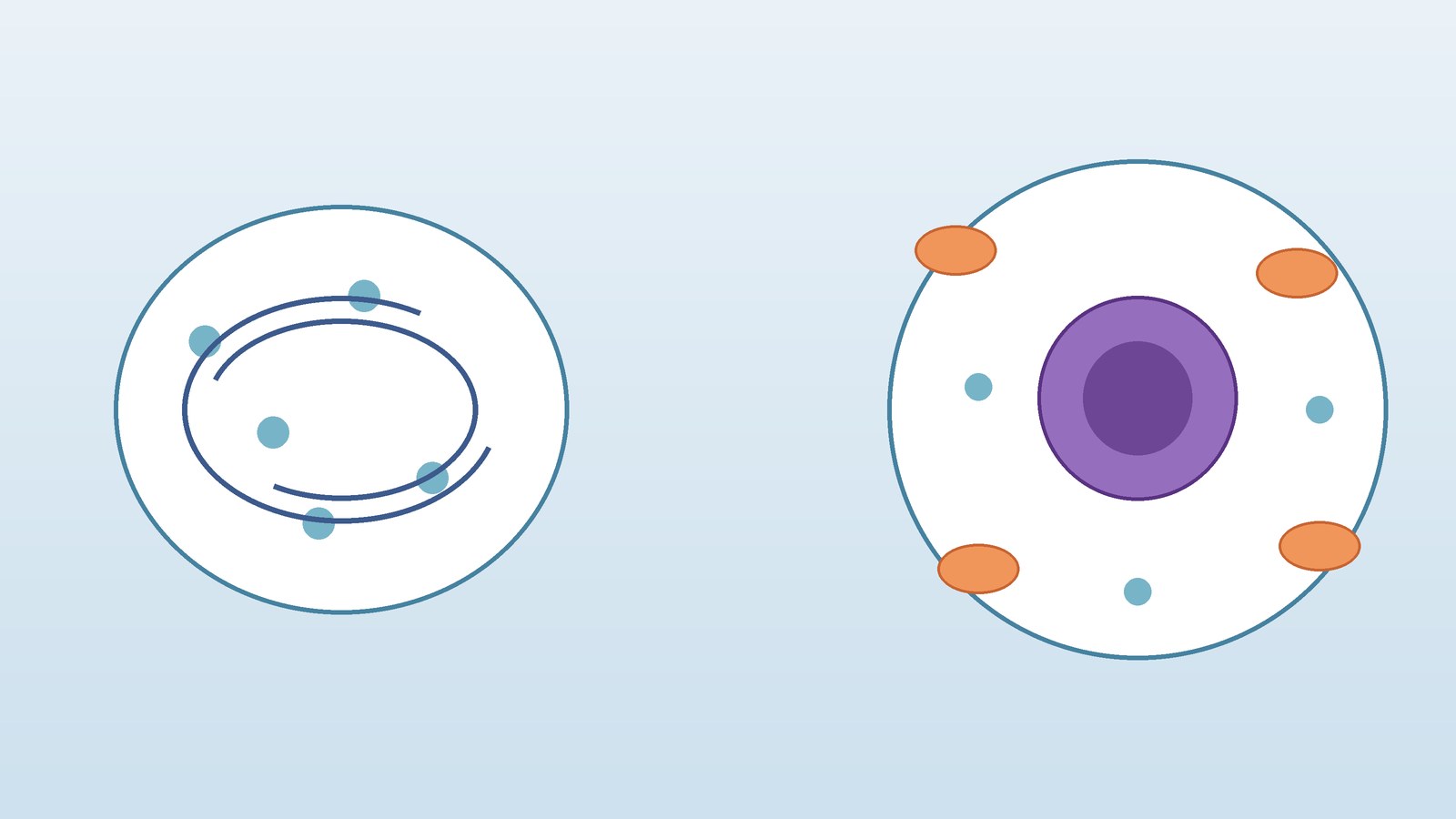
Short answer
- The main difference between prokaryotic and eukaryotic cells is how they organize their genetic material and their interior. A prokaryotic cell has no membrane bound nucleus and no membrane bound organelles. Its DNA sits in a region of the cytoplasm called the nucleoid. A eukaryotic cell has a membrane bound nucleus that holds its DNA, plus membrane bound organelles that divide the work of the cell into compartments. Prokaryotic cells belong to the organisms of the domains Bacteria and Archaea, and they are almost always single celled. Eukaryotic cells belong to animals, plants, fungi, and protists, and they may be single celled or part of a multicellular organism. Prokaryotic cells are also usually much smaller than eukaryotic cells, they divide by binary fission rather than by mitosis or meiosis, and they carry smaller 70S ribosomes instead of the larger 80S ribosomes found in the cytoplasm of eukaryotic cells.
The two categories of cells
Every living thing is made of cells, but not all cells are built the same way. Biologists sort cells into two broad categories: prokaryotic cells and eukaryotic cells. The sorting is not about what the organism looks like from the outside. It is about the internal architecture of the cell itself. A bacterium under a microscope looks nothing like a human skin cell, yet the deeper point is structural. One cell type keeps its DNA loose in the cytoplasm without a nucleus. The other encloses its DNA in a nucleus and adds a set of internal compartments.
That distinction matters because it shapes almost everything else about the cell: how large it can grow, how it controls which genes are active, how it makes and moves proteins, and how it divides. Once you understand the nucleus difference, the other differences start to make sense as consequences of two different ways of organizing life at the smallest scale.
The names themselves give the first clue. Prokaryotic comes from Greek roots meaning before the nucleus. Eukaryotic comes from roots meaning true nucleus. In other words, the vocabulary of cell biology already tells you where to look first: find the nucleus, or find its absence, and you have found the dividing line.
At a glance: prokaryotic versus eukaryotic cells
| Feature | Prokaryotic cell | Eukaryotic cell |
|---|---|---|
| Nucleus | Absent; DNA is in the nucleoid region | Present; DNA is enclosed in a membrane bound nucleus |
| Membrane bound organelles | Absent | Present, including mitochondria, endoplasmic reticulum, and Golgi apparatus |
| DNA form | Usually a single circular chromosome, plus small plasmids in many bacteria | Multiple linear chromosomes inside the nucleus |
| DNA packaging | Not wrapped around histone proteins in the same way as eukaryotic DNA | Wrapped around histone proteins into chromatin |
| Cell size | Typically 0.1 to 5.0 micrometers in diameter, following OpenStax Concepts of Biology | Typically 10 to 100 micrometers in diameter, following OpenStax Concepts of Biology |
| Ribosomes | 70S ribosomes | 80S ribosomes in the cytoplasm |
| Cell wall | Present in most; bacterial walls contain peptidoglycan | Present in plants and fungi, absent in animal cells; plant walls contain cellulose and fungal walls contain chitin |
| Division | Binary fission | Mitosis for growth and repair, meiosis for gamete formation in sexually reproducing organisms |
| Organization | Almost always unicellular | Unicellular or multicellular |
| Examples | Bacteria and archaea | Animals, plants, fungi, and protists |
Read the table as a summary of typical cells, not as a set of absolute rules with no exceptions. Biology is full of edge cases, and this article flags the important ones later. The typical pattern, however, is clear and reliable: prokaryotic cells are simpler and smaller, while eukaryotic cells are compartmentalized and larger.
What all cells share
Before the differences, it helps to see what prokaryotic and eukaryotic cells have in common, because the shared features show that both are versions of the same basic design. OpenStax Concepts of Biology lists four components shared by all cells. First, every cell has a plasma membrane, an outer covering that separates the interior of the cell from its surroundings and controls what passes in and out. Second, every cell has cytoplasm, the jellylike region inside the cell where other components are found. Third, every cell has DNA, the genetic material that carries instructions for the cell. Fourth, every cell has ribosomes, particles that synthesize proteins.
That shared foundation is worth remembering when the differences start to pile up. A prokaryotic cell is not a failed eukaryotic cell, and a eukaryotic cell is not simply a prokaryotic cell with extra parts glued on. Both are complete, working cells. They solve the same basic problems of life, staying separate from the environment, copying genetic instructions, and making proteins, but they solve them with different internal layouts.
The nucleus: the defining difference
The nucleus is the single most reliable way to tell the two cell types apart. A eukaryotic cell has a membrane bound nucleus. Its DNA is enclosed inside that compartment, separated from the rest of the cytoplasm by the nuclear envelope. Inside the nucleus, the DNA is organized into chromosomes, and the processes that read genetic instructions can be controlled in a more separated way.
A prokaryotic cell has no nucleus at all. Its DNA is found in a central part of the cell in a darkened region called the nucleoid. The word nucleoid is easy to confuse with nucleus, but the two are not the same. A nucleus is a compartment with a membrane. A nucleoid is a region where DNA is concentrated, without a surrounding membrane. In a prokaryotic cell, the DNA sits directly in the cytoplasm, in contact with the rest of the cell contents.
This difference affects how the cell manages its genetic material. In a eukaryotic cell, transcription, the copying of DNA into RNA, happens inside the nucleus, while translation, the building of proteins from RNA instructions, happens outside it, at ribosomes in the cytoplasm or on the rough endoplasmic reticulum. That physical separation gives the eukaryotic cell an extra layer of control. In a prokaryotic cell, transcription and translation can happen in the same space, without a membrane between them, which fits the simpler and faster style of prokaryotic life.
DNA: circular chromosomes, linear chromosomes, and packaging
DNA organization is the second major difference, and it follows directly from the nucleus difference. A prokaryotic cell typically carries a single circular chromosome. Many bacteria also carry plasmids, small separate circles of DNA that can hold extra genes, such as genes that help the cell survive a particular challenge. The chromosome itself is not enclosed in a membrane, and it is not packaged with histone proteins in the same organized way as eukaryotic DNA.
A eukaryotic cell carries multiple linear chromosomes inside its nucleus. Human cells, for example, carry many chromosomes rather than one circle of DNA. Eukaryotic DNA is wrapped around histone proteins to form chromatin, a packaging system that helps fit very long DNA molecules into the nucleus and helps control which genes are accessible at any given time. The combination of many linear chromosomes plus histone packaging is a hallmark of eukaryotic organization.
Two qualifications keep this picture honest. First, most bacterial chromosomes are circular, but not every prokaryotic chromosome follows that pattern, so the careful statement is usually, not always. Second, eukaryotic cells also carry small amounts of DNA outside the nucleus, in mitochondria and, in plants and algae, in chloroplasts. That extra DNA is a clue to the evolutionary history of those organelles, discussed later in this article. Neither qualification overturns the main pattern. It simply stops a useful summary from becoming an overstatement.
Organelles: compartments versus open floor plan
If the nucleus is the defining difference, organelles are the most visible difference. A eukaryotic cell contains membrane bound organelles, compartments with specialized functions. The mitochondria generate most of the cell chemical energy in the form of ATP. The endoplasmic reticulum helps make and move proteins and lipids. The Golgi apparatus modifies, sorts, and packages proteins for delivery. Lysosomes break down waste and worn out parts. In plant cells, chloroplasts carry out photosynthesis, and a large central vacuole stores water and helps maintain pressure. Each organelle is a room with a job, and the membranes between rooms let the cell run different chemical processes side by side without interference.
A prokaryotic cell has no membrane bound organelles. That does not mean it has no internal structure. It has ribosomes, a nucleoid, and often a cell wall, capsule, flagella, or pili. Some prokaryotes have internal membrane folds that increase surface area. But those folds are not separate membrane bound organelles in the eukaryotic sense, and they do not create the same system of compartments. The prokaryotic interior is closer to an open floor plan: everything shares the same cytoplasmic space.
Size: why eukaryotic cells can be larger
Size is one of the easiest differences to picture, and it is tied to the organelle difference. Following OpenStax Concepts of Biology as the single authority for the figures in this article, prokaryotic cells are 0.1 to 5.0 micrometers in diameter, while eukaryotic cells are 10 to 100 micrometers in diameter. In other words, a typical eukaryotic cell can be ten times wider or more than a typical prokaryotic cell, and far larger in volume.
Size figures vary between sources, so this point needs single authority labeling. Some educational sources give a narrower or wider range for prokaryotic cells, for example starting at about 1 micrometer rather than 0.1 micrometers, and some give a wider spread for eukaryotic cells. This article uses the OpenStax ranges above for every size statement, so that the comparison stays consistent. The direction of the comparison does not change between sources: prokaryotic cells are consistently described as significantly smaller than eukaryotic cells.
That is why size and complexity belong together in this comparison. A eukaryotic cell is not just a scaled up prokaryotic cell. It is a cell with a different internal system that makes larger size workable. In general, cell size is limited because volume increases much more quickly than surface area, so a larger cell has relatively less surface through which to take in materials and release wastes.
Ribosomes: same job, different size
Ribosomes are the exception that proves the shared foundation. Both prokaryotic and eukaryotic cells have ribosomes, because both need to make proteins. The ribosomes differ in size, measured in Svedberg units, abbreviated as S, which describe how fast particles settle during centrifugation rather than a simple physical diameter.
Prokaryotic ribosomes are 70S ribosomes. Eukaryotic ribosomes in the cytoplasm are 80S ribosomes. The textbook breakdown commonly given is that a 70S ribosome is made of a 50S large subunit and a 30S small subunit, while an 80S ribosome is made of a 60S large subunit and a 40S small subunit. The numbers do not add up in the simple way you might expect, because Svedberg units measure settling behavior, not mass alone, so 50 plus 30 does not equal 70 in that system.
The size difference has a practical consequence that students often meet in medicine: some antibiotics target the 70S ribosomes of bacteria while leaving the 80S ribosomes of human cells largely alone. That selective targeting is possible precisely because the two ribosome types differ. It is also a useful reminder that the 70S versus 80S distinction applies to the ribosomes in the main cytoplasm. The ribosomes inside mitochondria and chloroplasts resemble the 70S type, another clue to their origin, which is covered in the evolution section below.
Cell walls and outer layers: similar job, different materials
Cell walls are a good example of a feature that looks similar at first glance but differs in detail. Most prokaryotic cells have a cell wall. In bacteria, that wall is made of peptidoglycan, a mesh of sugars and amino acids. The wall gives the cell shape, protects it, and helps prevent dehydration. Many bacteria also have a polysaccharide capsule outside the wall, which helps the cell attach to surfaces. Archaea also have cell walls, but their wall chemistry differs from bacterial peptidoglycan, which is one of the reasons archaea are classified as a separate domain rather than grouped with bacteria.
Eukaryotic cell walls are a patchwork. Plant cells have walls made largely of cellulose. Fungal cells have walls made largely of chitin. Animal cells have no cell wall at all, relying on the plasma membrane and, in many tissues, an extracellular matrix for support. Protists vary: some have walls or shells, others do not. So the rule is not that one cell type has a wall and the other does not. The rule is that wall material and wall presence follow the organism group, and bacterial peptidoglycan is specific to bacteria.
Cell division: binary fission, mitosis, and meiosis
Cell division is where this comparison connects most directly to reproduction and growth, and it is also where this article deliberately stays brief, because the detailed account of prokaryotic division belongs to its own article.
Prokaryotic cells divide by binary fission. In binary fission, the cell copies its DNA and then splits into two daughter cells, each receiving a copy of the genetic material. The process is relatively simple and fast, which suits single celled organisms that multiply by making more copies of themselves. The companion article on binary fission covers the steps, the variations, and the limits of that process in detail, so this article treats it as the prokaryotic counterpart to the eukaryotic divisions below rather than repeating that full account.
Eukaryotic cells divide by mitosis for growth, repair, and asexual reproduction, producing daughter cells with the same chromosome number as the parent cell. Sexually reproducing eukaryotes also use meiosis to produce gametes, cells with half the chromosome number, so that fertilization restores the full number. Both mitosis and meiosis involve the nucleus, the organization of chromosomes, and a spindle apparatus, machinery that has no counterpart in binary fission. The complexity of eukaryotic division matches the complexity of the cell: when a cell has a nucleus and many linear chromosomes, dividing it safely takes more steps.
The key comparison, then, is not just speed. It is that prokaryotic division copies one circular chromosome and splits the cell, while eukaryotic division must copy, organize, and separate many chromosomes inside a nucleus, and then divide the cytoplasm and its organelles between the daughter cells.
Unicellular versus multicellular: one cell or many
Prokaryotic organisms are almost always unicellular. Each bacterium or archaeon is a single cell that carries out all the functions of life on its own. Some prokaryotes form chains, clusters, or biofilms, groups of cells living together, but each cell in the group remains a complete organism in its own right. There is no division of labor into tissues and organs.
Eukaryotic organisms may be unicellular or multicellular. Many protists, such as amoebas and paramecia, are single eukaryotic cells that live independently. Fungi include both single celled yeasts and multicellular molds and mushrooms. Animals and plants are multicellular, with cells specialized into tissues and organs. That range is possible because eukaryotic cells can specialize: a muscle cell, a leaf cell, and a nerve cell all share the same basic eukaryotic architecture, but they use it in different ways.
This difference helps explain why the two cell types dominate different parts of life. Prokaryotes excel as independent, fast multiplying, highly adaptable single cells. Eukaryotes excel when specialization and coordination pay off, from a single free living protist to a plant with roots, stems, and leaves. Neither strategy is better in general. Each wins in the environments it fits.
Examples that make the difference concrete
Examples help anchor the abstract comparison. On the prokaryotic side, Escherichia coli, the bacterium common in textbooks and laboratories, is a single cell with no nucleus, a circular chromosome, 70S ribosomes, and a peptidoglycan wall. Archaea found in hot springs, salty lakes, and other extreme environments are also prokaryotic, even though their biochemistry differs from bacteria in important ways. The shared point is the cell plan, not the habitat.
On the eukaryotic side, a human cheek cell is a eukaryotic animal cell: it has a nucleus, mitochondria, endoplasmic reticulum, Golgi apparatus, and 80S ribosomes, but no cell wall and no chloroplasts. A plant leaf cell is also eukaryotic, but it adds a cellulose wall, chloroplasts, and a large central vacuole. A yeast cell is a single celled fungus, eukaryotic in plan, even though it lives independently like a bacterium. A paramecium is a single celled protist that swims, feeds, and reproduces as one eukaryotic cell. The variety on the eukaryotic side is enormous, but the nucleus and organelles are the common thread.
How eukaryotic cells may have evolved: the endosymbiont story
One of the most interesting parts of this comparison is the explanation for why eukaryotic cells have mitochondria and chloroplasts at all. The widely supported explanation is endosymbiotic theory: early eukaryotic cells took in free living bacteria, and instead of digesting them, kept them as internal partners. Over time, those partners became mitochondria, the energy organelles, and, in the plant and algal lineages, chloroplasts, the photosynthetic organelles.
The evidence fits the story in several ways. Mitochondria and chloroplasts have their own circular DNA, separate from the nuclear DNA. They have ribosomes that resemble the 70S type found in prokaryotes rather than the 80S type in the eukaryotic cytoplasm. They are surrounded by double membranes, consistent with an engulfed cell keeping its own membrane inside a host membrane. And they divide in a way that resembles binary fission, on a schedule that is partly independent of the host cell nuclear division.
Exceptions that refine the rules
A good comparison should name its exceptions, because exams and real biology both test them. Here are the main ones, stated carefully.
First, not every eukaryotic cell keeps its nucleus for its whole life. Mature mammalian red blood cells, for example, lose their nucleus as they mature, which leaves more room for hemoglobin, the molecule that carries oxygen. They are still eukaryotic cells by origin and lineage, but a blood smear will not show a nucleus in them. The rule is about the typical cell plan, not about every cell at every stage of life.
Second, not every prokaryotic cell is tiny. A small number of unusually large bacteria exist that break the usual size range. They are exceptions precisely because they need special adaptations, such as unusual shapes or internal organization, to overcome the surface-area-to-volume limit described in the size section. Their existence does not change the typical range, but it stops the size rule from being stated as absolute.
Third, not every prokaryotic chromosome is a single circle. Most are, but some prokaryotes carry linear chromosomes or multiple chromosomes. The careful wording is usually, not always, and that wording is used throughout this article for that reason.
Fourth, mitochondria and chloroplasts break the simple statement that all eukaryotic DNA is in the nucleus. They carry their own DNA, as explained above. The accurate statement is that the main genome of a eukaryotic cell is in the nucleus, with small additional genomes in those organelles.
Fifth, some prokaryotes have internal membrane folds that can look like compartments under a microscope. Those folds increase surface area, for example for photosynthesis in cyanobacteria, but they are not membrane bound organelles in the eukaryotic sense. The distinction is between folds of one membrane system and a set of separate compartments with different jobs.
When you can say both the rule and the exception, you understand the biology instead of just memorizing a table.
Differentiation from related articles
This article is the comparison. It answers how prokaryotic and eukaryotic cells differ, side by side, across nucleus, DNA, organelles, size, ribosomes, walls, and division. It does not repeat the full step by step account of prokaryotic division, which is the subject of the companion article on binary fission. Readers who want the division process in detail should read that article next, while readers who want the comparison should stay here. Similarly, this article mentions plant cell structures, such as the cellulose wall, chloroplasts, and central vacuole, only as part of the eukaryotic side of the comparison. The detailed labeled account of the plant cell, with its diagram and part by part guide, is the subject of the companion article on the plant cell diagram. That article, not this one, is the place for the full labeled tour of a plant cell. Keeping the three articles separate avoids duplication: comparison here, division process in the binary fission article, and plant cell anatomy in the plant cell diagram article.
Frequently asked questions
What is the main difference between prokaryotic and eukaryotic cells?
The main difference is the nucleus and the organelles. Prokaryotic cells have neither a membrane bound nucleus nor membrane bound organelles, and their DNA sits in the nucleoid region. Eukaryotic cells have a membrane bound nucleus and membrane bound organelles, and their DNA sits inside the nucleus as multiple linear chromosomes.
Are prokaryotic cells always smaller than eukaryotic cells?
Typically, yes. Following OpenStax Concepts of Biology, the ranges given in this article do not overlap: prokaryotic cells are 0.1 to 5.0 micrometers in diameter and eukaryotic cells are 10 to 100 micrometers in diameter. A small number of unusually large bacteria break the usual prokaryotic range, so size is strong supporting evidence rather than an absolute rule on its own.
Do prokaryotic cells have a nucleus?
No. Prokaryotic cells have a nucleoid region where their DNA is concentrated, but they have no membrane bound nucleus. The nucleoid and the nucleus are different structures, and confusing them is one of the most common mistakes in this topic.
Do prokaryotic cells have organelles?
Prokaryotic cells have no membrane bound organelles, such as mitochondria, endoplasmic reticulum, or a Golgi apparatus. They do have ribosomes, which make proteins, and they may have a cell wall, capsule, flagella, or pili. The precise statement is no membrane bound organelles, not no internal structures at all.
What organisms have prokaryotic cells and what organisms have eukaryotic cells?
Bacteria and archaea have prokaryotic cells. Animals, plants, fungi, and protists have eukaryotic cells. That grouping follows the domains of life: Bacteria and Archaea are prokaryotic domains, while Eukarya is the eukaryotic domain.
How do prokaryotic and eukaryotic cells divide differently?
Prokaryotic cells divide by binary fission, copying their DNA and splitting into two daughter cells. Eukaryotic cells divide by mitosis for growth and repair, and sexually reproducing eukaryotes use meiosis to make gametes. The eukaryotic processes are more complex because they must organize and separate many chromosomes inside a nucleus.
Why do eukaryotic cells have mitochondria if prokaryotic cells do not?
Eukaryotic cells have mitochondria because, according to endosymbiotic theory, an early host cell took in bacteria that became permanent internal partners and then organelles. Mitochondria keep their own circular DNA and 70S-like ribosomes, which are traces of that bacterial origin. Prokaryotic cells never went through that partnership as hosts, so they have no mitochondria.
Is a virus a prokaryotic cell?
No. A virus is not a cell at all, prokaryotic or eukaryotic. Viruses have genetic material and, in some cases, a protein coat or envelope, but they have no cytoplasm, no ribosomes of their own, and no metabolism. They can only replicate inside a host cell. The prokaryotic versus eukaryotic comparison applies to cells, and viruses sit outside that comparison.
Which cell type came first?
Prokaryotic cells came first. Bacteria and archaea are the older cell plans, and eukaryotic cells evolved later, in part through the endosymbiotic partnerships described above. Eukaryotic complexity builds on the earlier prokaryotic foundation rather than replacing it, which is why both cell types still thrive today.
Sources & references
- OpenStax, Concepts of Biology, section 3.2, Comparing Prokaryotic and Eukaryotic Cells, Rice University. This is the single authority for the cell size ranges used in this article, and the source for the shared cell components, the nucleoid, the absence of a nucleus and membrane bound organelles in prokaryotic cells, the membrane bound nucleus and organelles in eukaryotic cells, bacterial peptidoglycan walls, capsules, flagella, and pili, and the surface area and volume explanation of cell size limits.
- OpenStax, Biology 2e, section 4.2, Prokaryotic Cells, Rice University. This supports the same prokaryotic cell plan, including the nucleoid, ribosomes, cell membrane, and cell wall as the structures present in all prokaryotes, with other structures present in some but not all bacteria.


