Short answer
- A small amount of clear fluid or light crust after sleep can be normal if it is stable from day to day, wipes away easily, and both eyes look clear and comfortable.
- Change matters more than a single morning crust. A sudden increase, a change in color or thickness, discharge in one eye only, or discharge with redness or squinting is the pattern to act on.
- Color gives clues, not a diagnosis. Clear and watery discharge often means extra tears. Thick white, gray, yellow, or green mucus can occur with dry eye, infection, inflammation, or a corneal problem, and those causes overlap.
- Dry eye is a common cause that is easy to mistake for a simple infection. In dry eye the eye does not make enough of the watery part of tears, and thick discharge returns soon after cleaning.
- Squinting, holding an eye closed, pawing at the face, cloudiness, and swelling are red flags for pain or injury. Do not wait to see whether those signs settle.
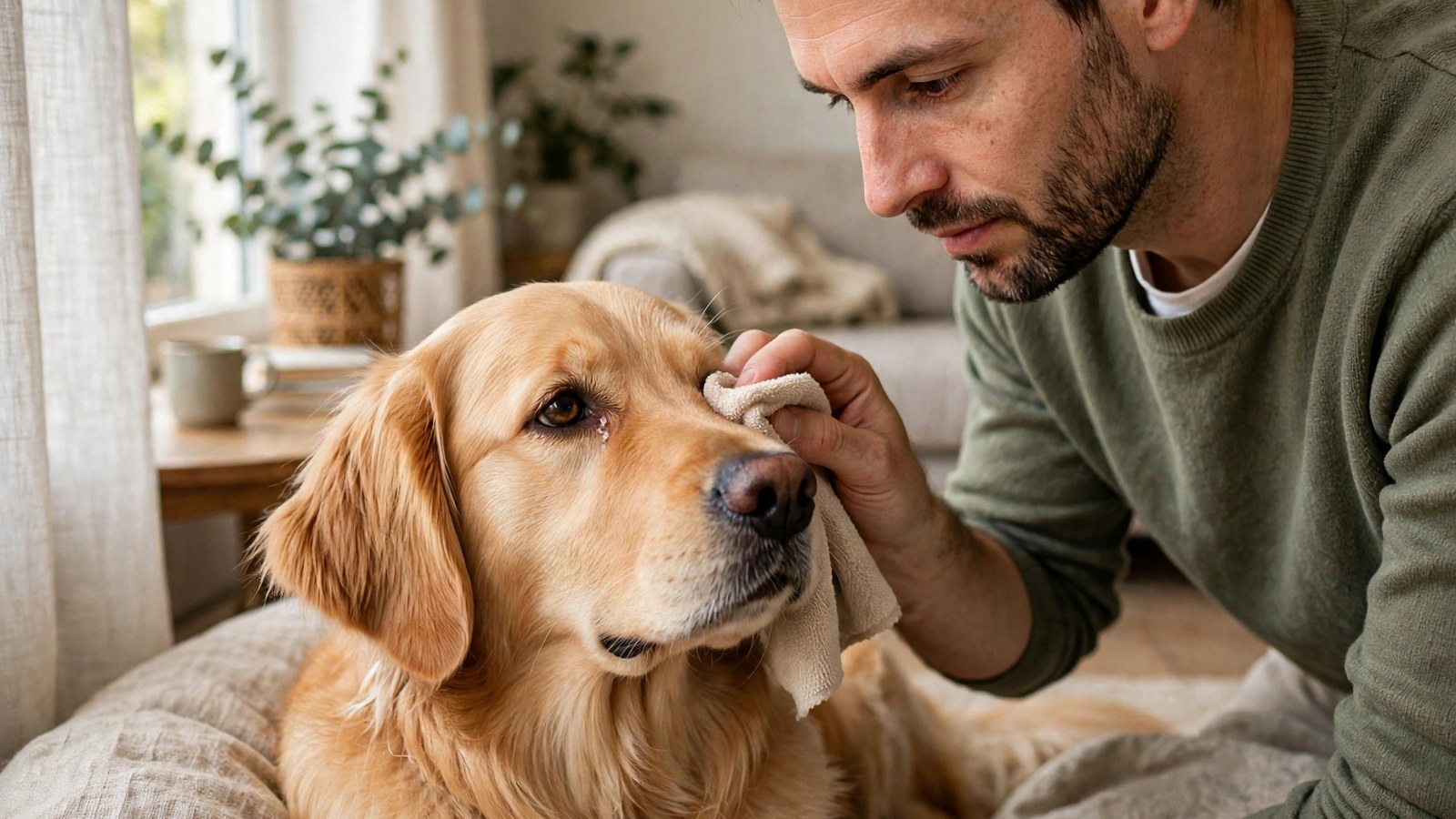
- At home, limit yourself to gentle cleaning of the fur around a comfortable eye with a clean, soft, damp cloth. Do not put leftover, human, or another dog's eye drops in the eye.
- A veterinarian finds the cause by examining the whole eye, which may include measuring tear production, staining the cornea, checking the eyelids and tear drainage, and measuring pressure inside the eye when indicated.
- This article is general information only, not a diagnosis. If discharge is abnormal, persistent, recurring, or paired with pain, arrange a veterinary examination promptly.
Introduction
A little clear fluid, or a small crust in the corner of the eyes after sleep, can be normal in a dog. Discharge becomes a concern when it is new, heavy, thick, yellow or green, keeps coming back, affects only one eye when both were clear before, or arrives with redness, squinting, swelling, cloudiness, or signs of pain. The color and texture alone cannot diagnose the cause, and the same appearance can occur in several different conditions.
This article explains what normal discharge looks like, how tears normally work and drain, what different colors and textures may suggest, the common causes a veterinarian considers, what you can safely do at home before an appointment, and which signs need prompt care. It is general information only. It does not diagnose any individual dog, and it does not prescribe treatment, medication, drops, or doses for a reader’s dog. Where treatment is mentioned, it is described only in general categories as something a veterinarian may consider after an examination.
If your dog holds an eye closed, squints repeatedly, paws at an eye, or has a cloudy, swollen, or visibly injured eye, contact a veterinarian promptly. Eye problems can worsen quickly, and a painful eye needs an examination even when there is very little discharge to see.
What Normal Eye Discharge Looks Like
Dogs make tears throughout the day. Tears keep the surface of the eye moist, help wash away dust and debris, and support clear vision. After sleep, tears that have collected and dried at the inner corner can leave a small amount of clear fluid or a light colored crust. Many owners call that material eye goo or sleep. Veterinarians sometimes use the word rheum for the normal material that tears, mucus, and debris leave behind. Rheum on its own is not a diagnosis.
A normal pattern has three features. The amount is small. The pattern is similar from day to day. And the eye underneath looks comfortable once the crust is wiped away, with no redness, swelling, squinting, or cloudiness. A dog with a normal pattern will usually open both eyes fully, blink normally, and show no interest in rubbing or pawing at the face.
What should prompt a second look is change in any of those features. Discharge that suddenly becomes heavier, thicker, or colored is different from a stable morning crust. So is discharge that appears in one eye only, returns within hours of cleaning, mats the fur during the day, or is paired with a wet face, reddened skin below the eye, or a change in behavior. When you describe the problem to a veterinary practice, those details about timing, amount, and whether one or both eyes are affected are more useful than the crust alone.
Breed and face shape also affect what an owner sees. Dogs with prominent eyes, shallow sockets, facial folds, or long hair around the eyes can show more visible tearing and staining even when the eye is comfortable. That does not make new or worsening discharge safe to ignore in those breeds. The baseline for that dog matters, and a change from it is the signal to report.
How Tears Normally Work and Drain
Understanding discharge is easier once the tear system itself is clear. Cornell University College of Veterinary Medicine describes the tear film as having three layers: a mucin layer next to the eye, a watery layer in the middle, and an oily layer on the outside. Each layer has a different job. The mucin layer helps the film spread and stick to the surface. The watery layer, made by the tear producing glands, provides most of the moisture and washes the surface. The oily layer, made by small glands in the eyelids, slows evaporation so the film does not dry out between blinks.
Problems can arise from any layer. A shortage of the watery layer is the classic form of dry eye discussed below. Problems with the oily or mucin layers can also leave the surface poorly protected, even when watery tears look adequate. That is one reason a veterinarian looks at tear quality and the eyelids as well as tear quantity.
Tears also need somewhere to go. After washing the surface, tears normally drain through small openings near the inner corner of the eyelids and then through a duct toward the nose. If drainage is reduced, if the eyelid position keeps tears from reaching those openings, or if the eye makes more tears than the system can carry away, tears spill onto the face. The medical word for that overflow is epiphora. Overflow leaves the fur below the eye persistently wet, and tears left on light colored fur can stain it reddish brown. The staining pigment in tears is often described as porphyrin. Staining by itself is a sign of overflow, not a diagnosis of why the overflow is happening.
What the Color and Texture May Suggest
Color and texture help you describe what you see, and a veterinary team will ask about them. They do not identify a cause on their own, because different conditions produce overlapping appearances.
| What you see | What it may suggest in general terms | What it does not prove |
|---|---|---|
| Clear and watery | Extra tears from irritation, wind, dust, allergy, pain, a foreign object, or poor drainage | That the eye is healthy, or that allergy is the cause |
| White or gray mucus | Mucus collecting when the watery part of tears is too low, as in dry eye, or with conjunctival inflammation | That a simple infection is present |
| Yellow or green, thick | Inflammation or infection, dry eye, a corneal wound, or inflammation inside the eye | That antibiotic drops alone will fix the problem |
| Small light crust after sleep only | Dried tears and mucus, often normal when stable and the eye is comfortable | That a new or heavier crusting pattern is also normal |
| Thick crust sealing the lids, or crust returning heavily in the day | Ongoing discharge that needs examination, especially with redness or squinting underneath | That pulling the crust away has treated anything |
| Red eye with any discharge | Inflammation or irritation of the eye or eyelids, with many possible causes | Which of those causes is present in your dog |
Clear and watery
A constantly wet face, even with clear fluid, deserves a check. Ongoing tearing can irritate the skin below the eye, keep the fur damp, and stain it. Clear tearing that starts suddenly in one eye, or that comes with blinking or squinting, also deserves prompt attention, because pain and a foreign object or corneal injury can all drive extra tear production.
White or gray mucus
Thick white or gray mucus that returns soon after cleaning is a pattern veterinarians associate in particular with dry eye. The eye may also look red or dull, and the dog may blink or squint more than usual. Because the discharge of dry eye can look like the discharge of an infection, the two are easily confused at home. Measuring tear production is what separates them in the clinic.
Yellow or green discharge
Yellow or green discharge can occur with infection and with inflammation that has no simple bacterial cause. Dry eye, a corneal ulcer, and inflammation inside the eye can all produce colored discharge. That overlap is why a veterinarian usually needs to examine the eye, check the cornea, and measure tears before choosing any treatment category, and why guessing from color alone is risky.
Crusty eyes
Judge crust by what lies underneath it and by how it behaves. A small morning crust over a comfortable eye that stays stable is a different picture from thick crust that seals the eyelids shut, returns heavily during the day, or covers a red and sore eye. Do not pull hard crust away by force. Soften it first with a clean, soft, damp cloth held gently against the closed lid, and arrange care if the eye underneath looks sore.
Common Causes of Dog Eye Discharge
Irritation and foreign objects
Dust, wind, smoke, pollen, shampoo, and grooming products can irritate the surface and cause watering. A grass seed, a hair, grit, or another small object can lodge on the surface or under an eyelid and do the same. One eye affected, sudden onset, and signs such as blinking, squinting, or rubbing the face all raise concern for a local cause of this kind. A foreign object can scratch the cornea even when nothing is visible to the owner. Do not try to remove material that appears stuck to the surface of the eye. That is work for a veterinary examination, with the eye properly lit and the dog safely restrained.
Allergies and conjunctivitis
The conjunctiva is the thin membrane that lines the inside of the eyelids and covers the white of the eye at the front. The Merck Veterinary Manual notes that it helps make tears, protects the eye, supports eye movement, and helps the cornea heal after injury, which is why problems there matter beyond simple redness.
Conjunctivitis means inflammation of that membrane. Merck describes its signs as redness, swelling of the tissue around the cornea, discharge, and mild discomfort, and stresses that the appearance of the conjunctiva alone is usually not enough for a veterinarian to diagnose the cause from a physical examination alone. History, testing of conjunctival samples in some cases, and a tear test may all be needed.
Environmental allergies and irritants are common causes, and allergic eyes often come with itching, licking, or ear irritation elsewhere on the body. But watery discharge alone does not confirm an allergy, especially when only one eye is affected or the dog seems uncomfortable. Merck notes that conjunctivitis in only one eye may result from a foreign object, inflammation of the tear sac, or dry eye, while conjunctivitis in both eyes is commonly caused by a viral or bacterial infection, with irritants and allergens as other common causes. Treating redness and discharge as if the cause were already known can therefore miss the foreign object, the drainage problem, or the tear shortage that is actually driving it.
Dry eye
Dry eye, called keratoconjunctivitis sicca or KCS, occurs when there is inadequate production of the watery component of tears, in Cornell’s wording. Cornell states that the most common cause is immune mediated inflammation of the tear producing glands, in which the immune system damages those glands. Other causes Cornell lists include chronic allergies, endocrine diseases such as diabetes and hypothyroidism, congenital factors, surgical removal of the third eyelid gland, infections such as canine distemper virus, certain drug toxicities, and radiation treatment. A less common form follows loss of nerve function to the tear gland, and that form often affects one eye only, sometimes with dryness of the nostril on the same side.
Signs Cornell lists include thick yellow green discharge, a dull or dry appearance to the eye, redness, squinting, and pawing or rubbing at the face, with blood vessels or dark pigment appearing on the cornea in chronic cases. KCS typically affects both eyes. Without management, Cornell warns, the condition can lead to scarring, blood vessel formation, and secondary infection on the cornea, and potentially to permanent vision loss, and affected dogs are at risk of painful corneal ulcers.
Breed predisposition is part of the picture. Cornell lists the American Cocker Spaniel, English Bulldog, Lhasa Apso, West Highland White Terrier, Cavalier King Charles Spaniel, Shih Tzu, and Pug as breeds in which KCS is more common, while noting it can occur in any breed and is most common in middle aged dogs. Owners of those breeds should treat recurring thick discharge as a reason to ask specifically about tear testing, rather than as a routine nuisance to keep wiping away.
Diagnosis is by measurement, not by appearance. Cornell describes the Schirmer tear test as measuring watery tear production over one minute, using a small strip placed in the eye. A veterinarian may also stain the cornea to look for ulcers that have developed alongside the tear shortage. In general terms, treatment categories a veterinarian may consider include medicated drops intended to increase natural tear production and reduce inflammation, lubricating preparations, and treatment for any secondary infection present. Cornell notes that most dogs respond well, that improvement may take a few weeks and occasionally up to twelve weeks, and that KCS usually requires lifelong management. Those are categories to discuss with your own veterinarian, not a plan to copy at home.
Corneal scratches and ulcers
The cornea is the clear surface at the front of the eye. It can be scratched during play, by a branch, a thorn, or a grass seed, by another animal, or by the dog rubbing an already irritated eye. When the surface layer is lost, the defect is called a corneal ulcer. Ulcers can cause excessive tearing, watery or colored discharge, redness, sensitivity to light, blinking, and squinting.
Two points matter for owners. First, squinting is a sign of discomfort even when discharge looks mild, so a quiet looking eye that is held half closed is not a reassuring picture. Second, ulcers can deepen and threaten vision, and some eye drops that help other conditions can worsen an ulcer. That is why a veterinarian stains the cornea before choosing drops for a painful eye, and why a painful or tightly closed eye should be checked promptly rather than treated with whatever is already in the house.
Blocked tear ducts and poor drainage
When the drainage route is blocked, narrowed, not fully open from birth, inflamed, or pressed on by nearby tissue, tears spill onto the face instead of draining toward the nose. The result is persistent wet fur, reddish brown staining, and sometimes sore skin below the eye where dampness sits. Inflammation of the tear sac can also cause watering and conjunctivitis that keeps returning.
Facial structure plays a role. Dogs with short muzzles, shallow sockets, or prominent eyes may be more prone to overflow, and eyelid position or hair near the drainage opening can add to it. Persistent watery eyes still deserve evaluation even in those breeds, because irritation, eyelid problems, and corneal pain produce the same wet face.
Eyelid and eyelash problems
Eyelids that roll inward, a condition called entropion, bring fur into contact with the surface. Eyelids that roll outward, extra eyelashes growing from abnormal places, or single misplaced lashes can also rub the cornea. The result is recurring watering, redness, discharge, and, over time, corneal wounds. Hair in facial folds or long hair falling across the eye can make the area harder to keep clean and can add to irritation.
Repeated wiping will not correct a lid or lash that keeps touching the eye. The eyelids, lashes, and surrounding skin need to be examined, sometimes with magnification and with the eyelid turned to check its inner surface, so that any correction addresses the structure causing the problem rather than the discharge it produces.
Other eye conditions and wider illness
Discharge sometimes accompanies a condition that sits deeper than the surface. Raised pressure inside the eye, called glaucoma, and inflammation inside the eye, called uveitis, can both cause redness, tearing, cloudiness, a pupil that looks different, and pain. Certain infections and illnesses affecting the whole body can also show up in the eyes. These possibilities matter most when there is a sudden change in vision, pupils that look unequal, a visibly larger or cloudier eye, marked swelling, or signs of illness elsewhere.
Discharge paired with lethargy needs particular care. A dog that has mucus on the eyes and is also unusually tired, uninterested in food or walks, or reluctant to move should prompt a veterinary call the same day. That combination can mean the problem is not limited to the eye, or that the dog is in enough discomfort to change its behavior. Do not assume a tired dog with goopy eyes simply needs rest.
Owners sometimes notice discharge after a vaccination visit. Mild, brief tearing can follow stress, handling, or dust in the clinic, but eye discharge is not a typical expected effect of vaccination itself. If it persists, worsens, or comes with redness, squinting, swelling, or lethargy, call your veterinarian, mention the timing, and do not delay an eye examination because the visit seems like an explanation.
When to Call a Veterinarian
Some signs should not wait for a routine appointment. Seek veterinary attention right away, including from an emergency service if your usual practice is closed, if your dog:
- Holds an eye closed, squints persistently, or paws or rubs at an eye
- Has a cloudy, blue looking, or visibly injured eye, or something appears embedded in the eye
- Shows obvious eye pain, sudden swelling of the eye or eyelids, or a sudden change in vision
- Has had a suspected scratch, a blow to the eye, or any chemical getting into the eye
Contact your veterinarian promptly, and describe what you see, if you notice any of the following:
- Yellow or green discharge, or thick mucus that repeatedly returns after cleaning
- Redness or swelling of the eye or eyelids, especially if one eye is much worse than the other
- A sudden increase in tearing, particularly in one eye
- One eye that looks different from the other, or pupils that look unequal
- Persistent wetness, staining, or irritated skin below the eye
- Discharge paired with lethargy, poor appetite, vomiting, or other signs of illness
- Discharge that seems to come from a swollen or painful area above or below the eye, rather than from the eye itself
Finally, contact your veterinarian if unusual discharge persists or keeps returning, even if your dog otherwise seems well. A recurring pattern often means an underlying cause such as dry eye, a drainage problem, or a lid problem has not yet been found.
What You Can Do at Home Before the Appointment
Home care for eye discharge is deliberately narrow. It supports a comfortable eye while you arrange care. It does not treat causes.
If the eyes look comfortable and you see only minor discharge on the surrounding fur, you may gently wipe the buildup away with a clean, soft cloth dampened with warm water. Keep pressure light, work on the fur and the closed lid rather than the eyeball itself, use a fresh part of the cloth for each eye, and wash your hands before and after. Soften any crust first rather than pulling it free. Stop and arrange care if cleaning reveals redness, swelling, or soreness underneath, or if your dog resists because the eye hurts.
If a chemical gets into the eye, flush the eye right away with clean water or sterile saline for several minutes, then contact a veterinarian for prompt evaluation. Do not wait to see whether the eye settles after a chemical exposure.
Useful notes help the veterinary team more than repeated cleaning does. Record when the discharge started, whether one or both eyes are affected, the color and thickness, and whether you have noticed squinting, rubbing, redness, swelling, or lethargy. A clear photograph taken before you clean the eye can show the team what the eye looked like at its worst.
The limits matter as much as the steps. Do not put leftover eye drops, human eye medication, or drops prescribed for another dog or for an earlier problem into the eye without veterinary advice. Eye medicines are chosen for a specific diagnosed problem, a veterinarian may need to check for a corneal ulcer first, and some drops can worsen an ulcer. Do not use home preparations, herbal washes, or tear stain products inside the eye. If something appears embedded in the eye, leave it in place and seek care promptly rather than trying to lift it out.
What a Veterinarian May Do
A veterinary eye examination looks at the whole system that produces discharge, not only at the discharge itself. It may include looking closely at the eyelids and lashes, the conjunctiva, the tear film, the drainage openings, and the cornea, often with magnification and a focused light.
Several tests are commonly described by veterinary authorities. The Schirmer tear test measures watery tear production over one minute with a small paper strip placed under the lower eyelid, and Merck notes that it is part of working out the cause of conjunctivitis as well as dry eye. Fluorescein staining places a dye on the surface that highlights scratches and ulcers so damage that is hard to see becomes visible. A veterinarian may also measure pressure inside the eye when glaucoma or inflammation inside the eye is a concern, check how tears drain, and, in some cases, examine samples taken from the conjunctiva.
Treatment then follows the cause found. In category terms only, a veterinarian may consider removing a foreign object, managing dry eye over the long term, treating inflammation or infection, addressing allergy, managing a blocked drainage route, correcting an eyelid or lash problem, or referring the dog to a veterinary ophthalmologist for complex or non healing problems. Cleaning the fur, even daily, does not substitute for any of that.
Frequently Asked Questions
Are goopy eyes normal in dogs?
A small amount of light crust after sleep can be normal if the eyes are clear and comfortable and the pattern is stable. Heavy, thick, colored, or rapidly returning discharge, or goopy eyes paired with redness, squinting, swelling, or pawing at the face, should be examined. A large amount of discharge that keeps coming back is not a normal pattern to ignore, even in a breed that usually shows some tearing.
Does green discharge always mean infection?
No. Green discharge can appear with infection, but Cornell notes that thick green discharge is also a hallmark sign of dry eye, and corneal wounds and inflammation inside the eye can produce colored discharge as well. An examination, usually including a tear test and corneal staining, is needed to find the underlying cause before treatment is chosen.
Can allergies cause watery eyes in dogs?
Yes. Environmental allergies and irritants are common causes of conjunctivitis and tearing, as Merck describes, and allergic signs often appear in both eyes alongside itching or ear irritation. Watery eyes can also be a response to pain, a foreign object, a corneal ulcer, or poor tear drainage, so persistent or one sided symptoms deserve an examination rather than an assumption about allergy.
Can I clean my dog’s eye discharge with warm water?
You can gently clean discharge from the fur around a comfortable looking eye with a soft cloth dampened with warm water, using light pressure and a fresh part of the cloth for each eye. Do not rub the eyeball itself, do not pull hard crust free without softening it, and do not try to remove anything stuck to the eye. Cleaning is supportive only. It does not replace veterinary care when discharge is abnormal, colored, thick, or recurring.
Will tear stains go away if I clean them every day?
Regular gentle cleaning can keep the fur and skin below the eyes more comfortable. Staining itself may return for as long as tears keep overflowing onto the face, because the pigment is in the tears. A veterinarian can check whether irritation, drainage, eyelid position, or facial structure is contributing, and whether any of those causes can be addressed.
How quickly should a squinting eye be seen?
Promptly, and the same day where you can arrange it. Squinting and holding an eye closed are signs of discomfort that can accompany a corneal scratch or ulcer, a foreign object, dry eye, or pressure or inflammation inside the eye. Those causes cannot be separated at home, and some worsen quickly. If your usual practice is closed and the eye is painful, cloudy, swollen, or injured, use an emergency service rather than waiting.
Sources & references
- Merck Veterinary Manual, Dog Owners edition. "Disorders of the Conjunctiva in Dogs." Describes the role of the conjunctiva, the signs of conjunctivitis, why appearance alone does not establish the cause, the role of history, conjunctival testing, and the Schirmer tear test, and common causes in one eye versus both eyes.
- Cornell University College of Veterinary Medicine, Richard P. Riney Canine Health Center. "Keratoconjunctivitis sicca (KCS) in dogs." Describes inadequate production of the watery component of tears, the three layers of the tear film, immune mediated and other causes, breed predispositions, clinical signs including thick yellow green discharge, diagnosis by Schirmer tear test and fluorescein staining, treatment categories, and lifelong management.


